Buscar en
Revista Española de Medicina Nuclear e Imagen Molecular (English Edition)
Toda la web
Inicio Revista Española de Medicina Nuclear e Imagen Molecular (English Edition) From interventionist imaging to intraoperative guidance: New perspectives by com...
Información de la revista
Vol. 37. Núm. 1.
Páginas 28-40 (Enero - Febrero 2018)
Compartir
Compartir
Descargar PDF
Más opciones de artículo
Visitas
12
Vol. 37. Núm. 1.
Páginas 28-40 (Enero - Febrero 2018)
Original article
From interventionist imaging to intraoperative guidance: New perspectives by combining advanced tools and navigation with radio-guided surgery
De la imagen intervencionista a la guía intraoperatoria: nuevas perspectivas combinando herramientas avanzadas y navegación con la cirugía radioguiada
Visitas
12
S. Vidal-Sicarta,
Autor para correspondencia
svidal@clinic.cat

Corresponding author.
, R. Valdés Olmosb,c,d, O.E. Niewege,f, R. Faccinig,h, M.R. Grootendorsti, H.J. Westerj, N. Navabk, B. Vojnovicl, H. van der Poelm, S. Martínez-Románn, J. Klodeo, F. Wawroschekp, F.W.B. van Leeuwenb
a Nuclear Medicine Department, Hospital Clínic Barcelona, Barcelona, Spain
b Interventional Molecular Imaging Laboratory, Department of Radiology, Leiden University Medical Centre, Leiden, The Netherlands
c Nuclear Medicine Section, Department of Radiology, Leiden University Medical Centre, Leiden, The Netherlands
d Department of Nuclear Medicine, Diagnostic Oncology Division, The Netherlands Cancer Institute – Antoni van Leeuwenhoek Hospital, Amsterdam, The Netherlands
e Melanoma Institute Australia, North Sydney, New South Wales, Australia
f Central Medical School, The University of Sydney, Sydney, New South Wales, Australia
g Physics Department, University of Rome “La Sapienza”, Rome, Italy
h IFNF Roma, Rome, Italy
i Lightpoint Medical Ltd., Chesham, Bucks, United Kingdom
j Chair of Pharmaceutical Radiochemistry, Technical University Munich, Munich, Germany
k Institute of Informatics, Technical University of Munich, Munich, Germany
l Department of Oncology, Cancer Research UK and Medical Research Council, Oxford Institute for Radiation Oncology, University of Oxford, Oxford, United Kingdom
m Urology Department, Netherlands Cancer Institute – Antoni van Leeuwenhoek Hospital, Amsterdam, The Netherlands
n Obstetrics and Gynaecology Department, University Hospital Germans Trias i Pujol, Badalona, Barcelona, Spain
o Clinic for Dermatology, University Hospital Essen, Essen, Germany
p Urology Department, Oldenburg Clinic, Oldenburg, Germany
Ver más
Este artículo ha recibido
Información del artículo
Resumen
Texto completo
Bibliografía
Descargar PDF
Estadísticas
Figuras (10)
Mostrar másMostrar menos
Abstract

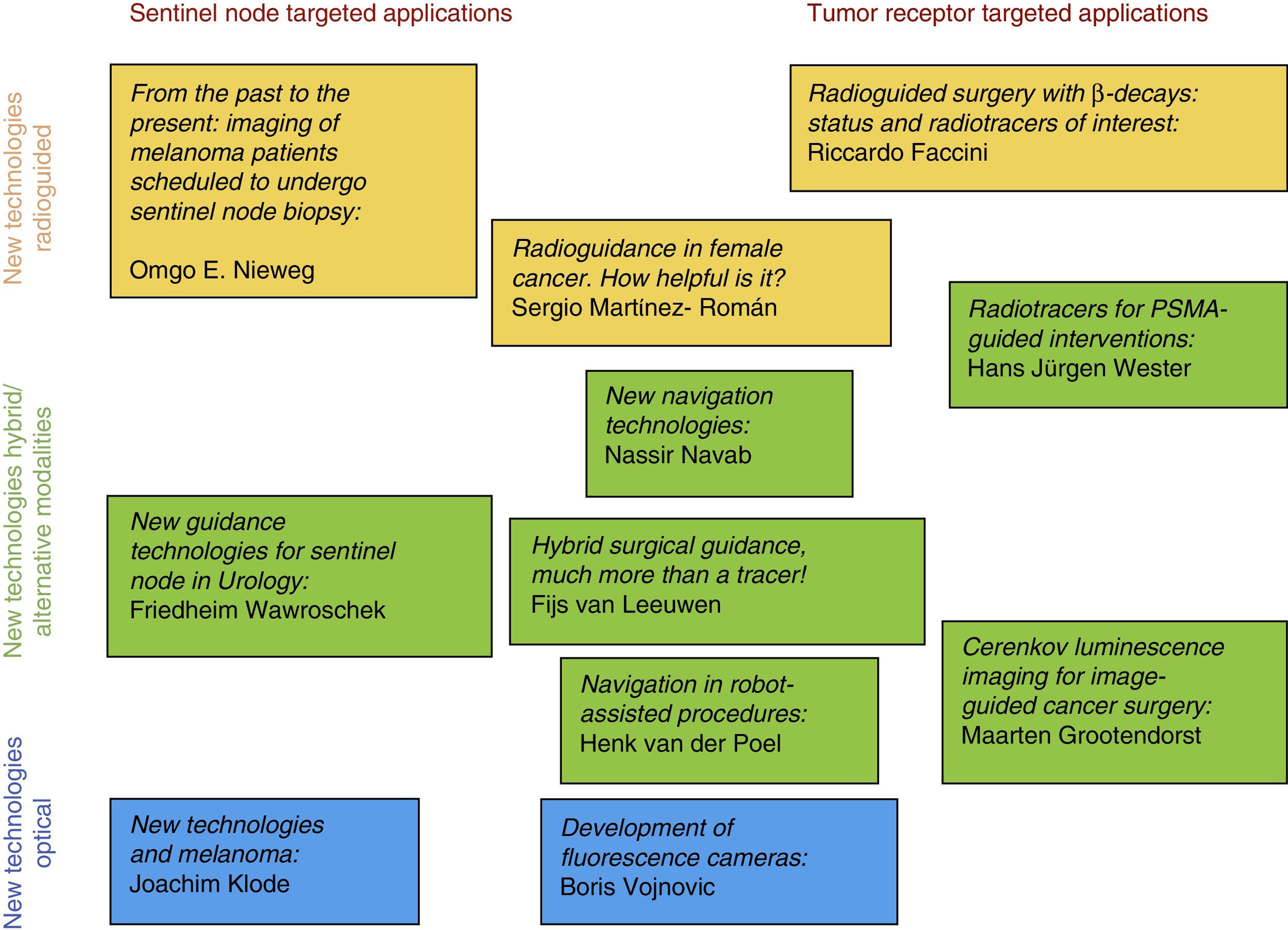
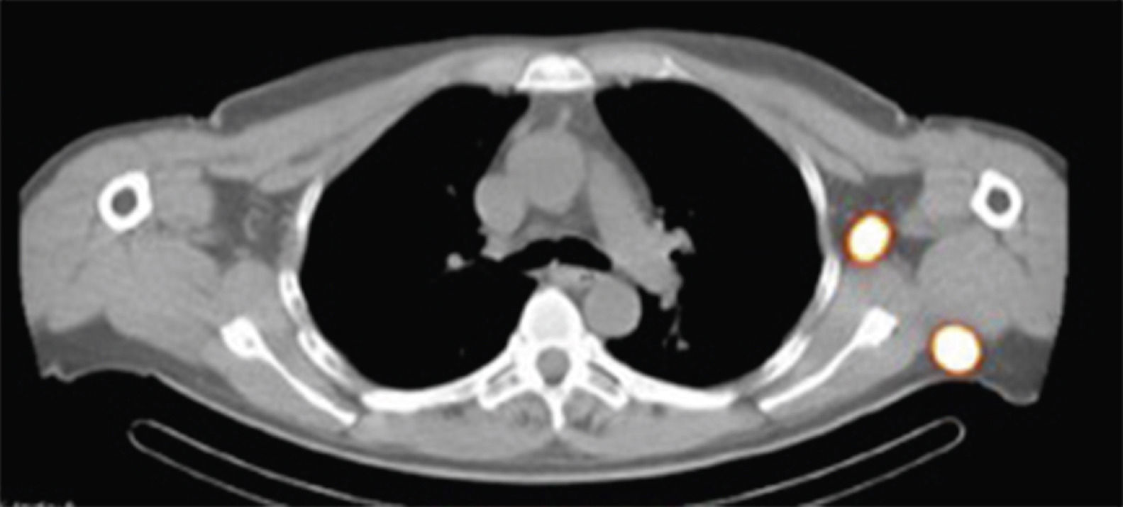
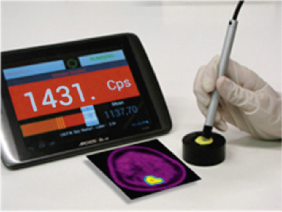
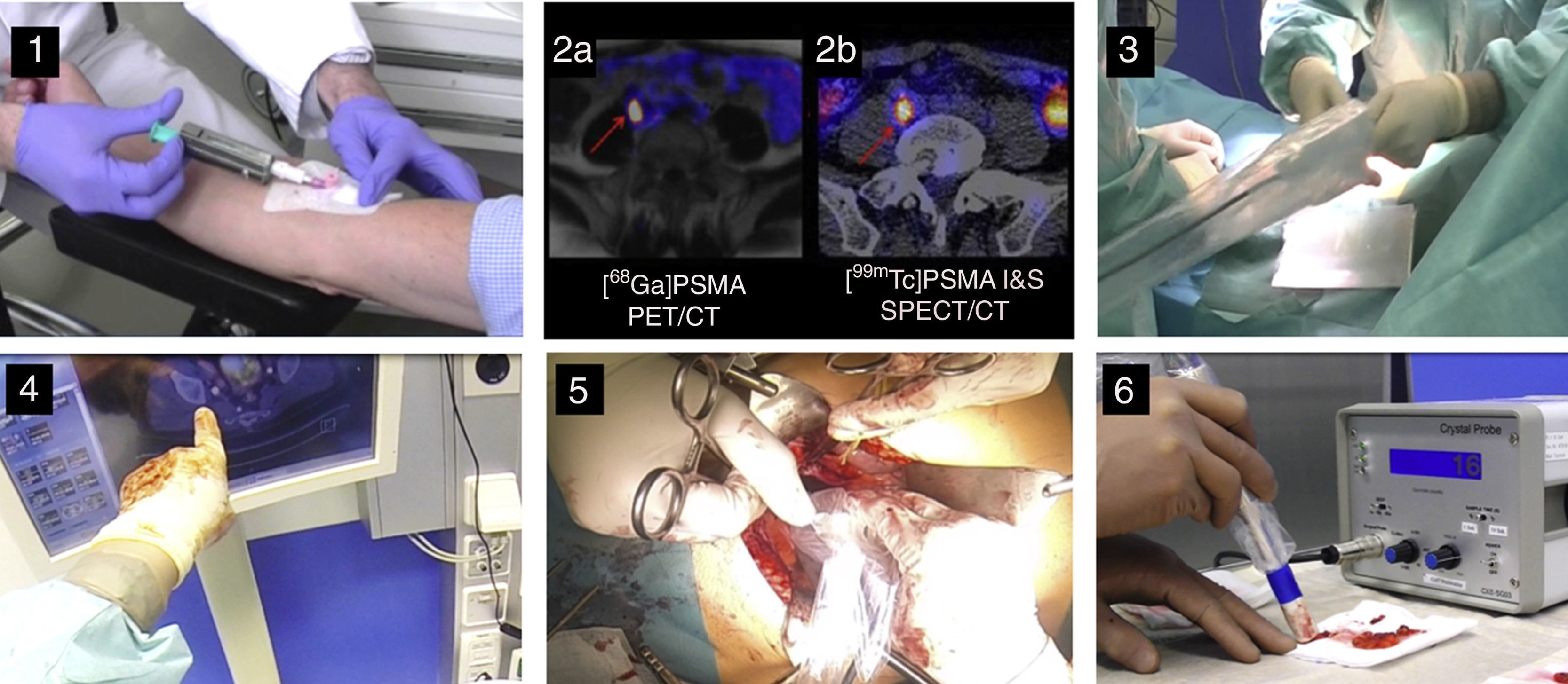
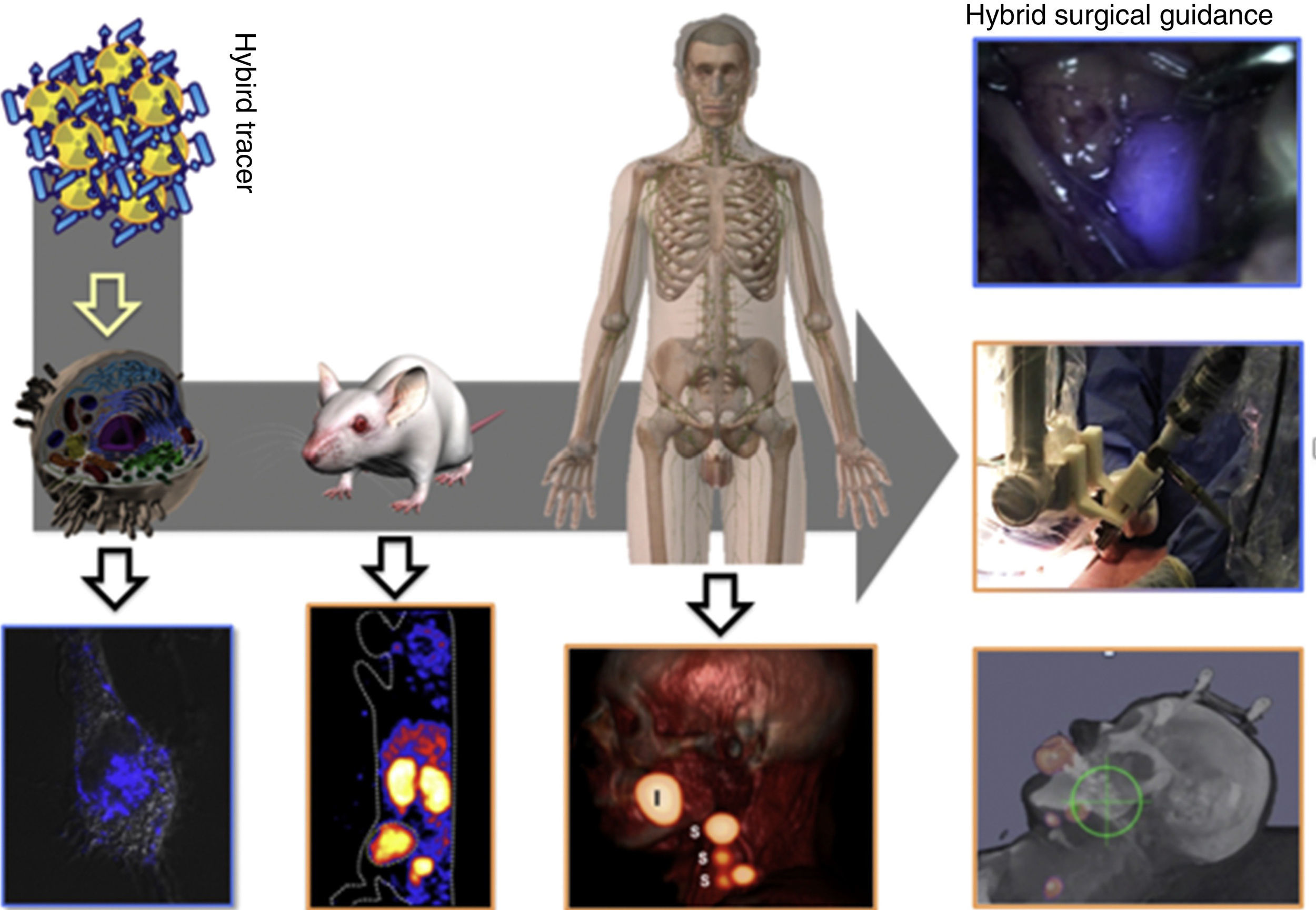
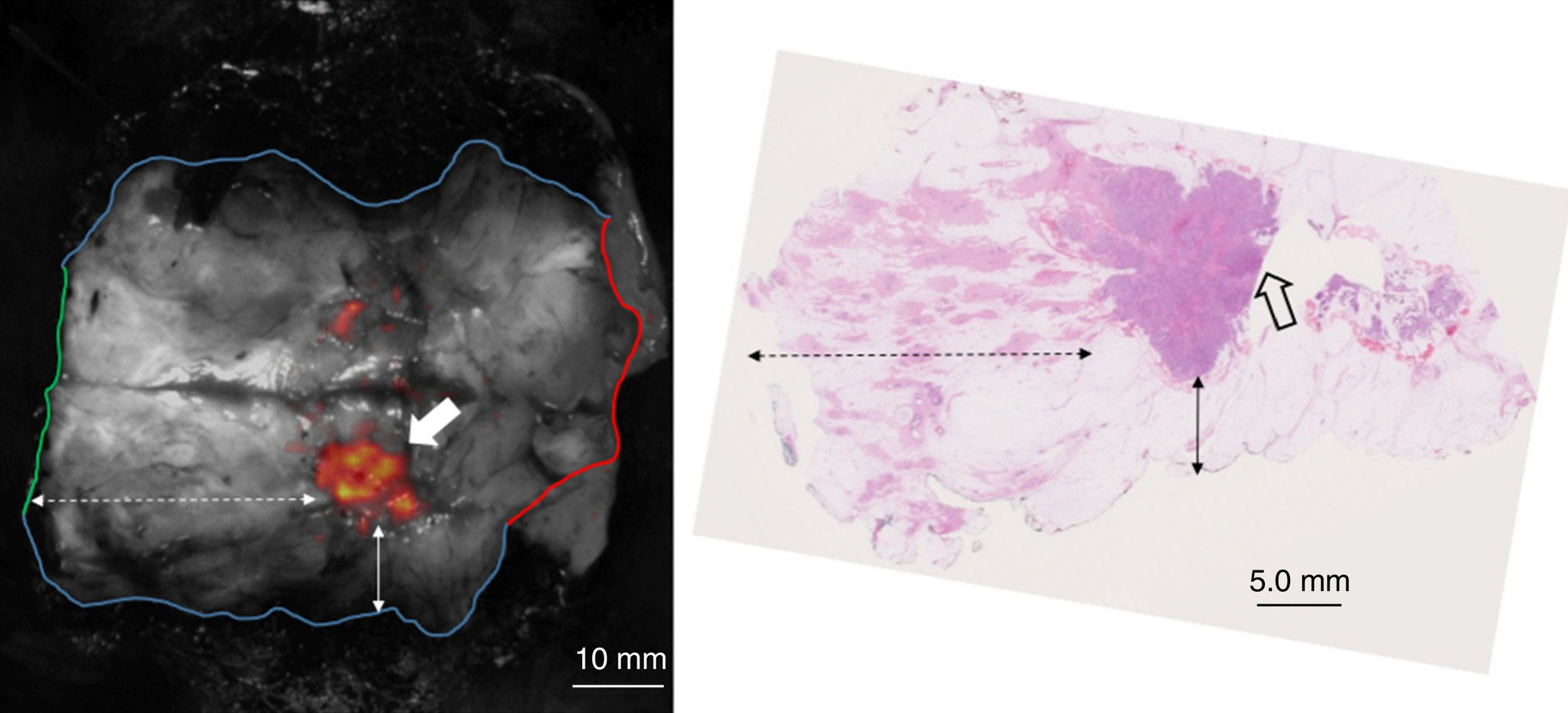
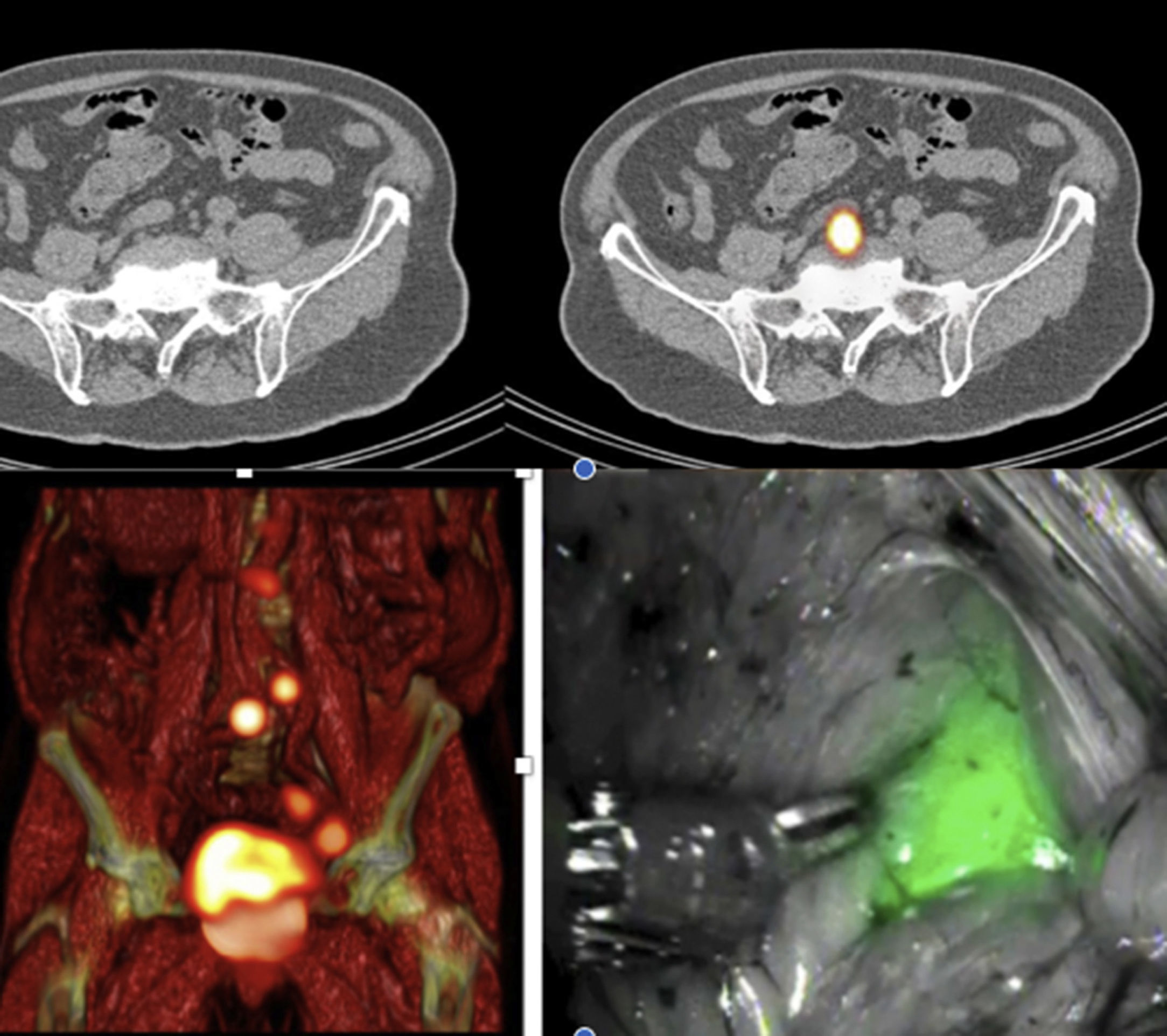
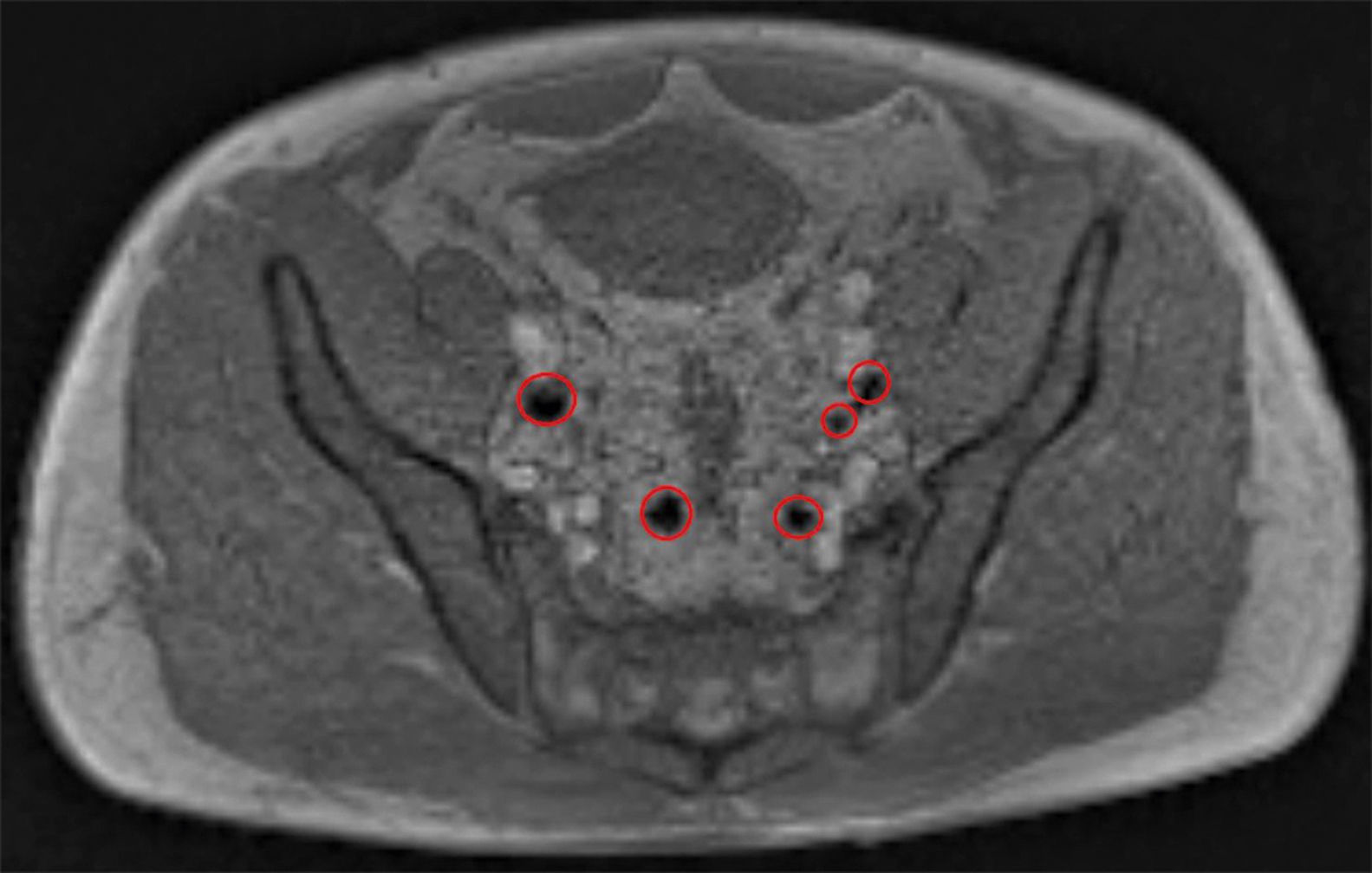
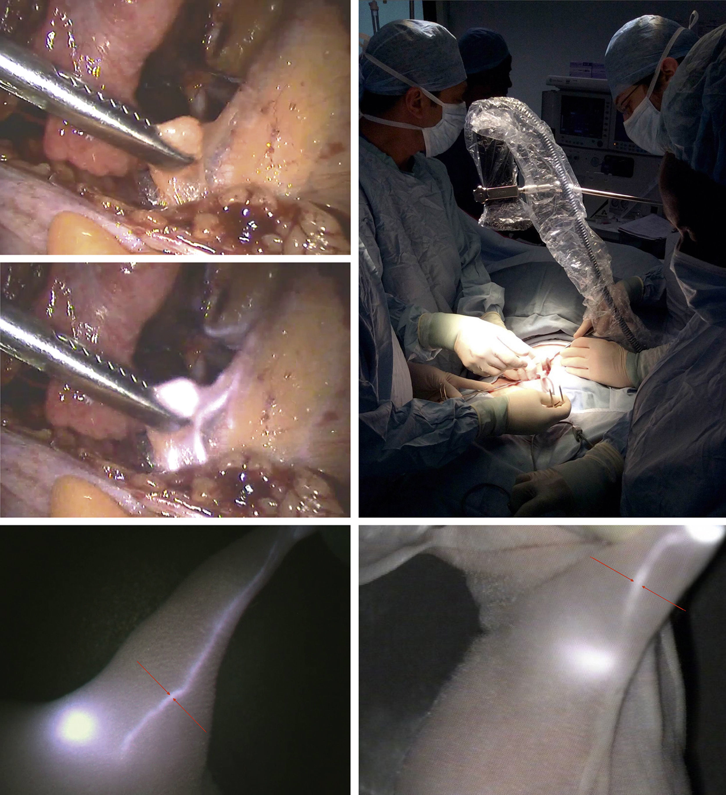
The integration of medical imaging technologies into diagnostic and therapeutic approaches can provide a preoperative insight into both anatomical (e.g. using computed tomography (CT), magnetic resonance (MR) imaging, or ultrasound (US)), as well as functional aspects (e.g. using single photon emission computed tomography (SPECT), positron emission tomography (PET), lymphoscintigraphy, or optical imaging). Moreover, some imaging modalities are also used in an interventional setting (e.g. CT, US, gamma or optical imaging) where they provide the surgeon with real-time information during the procedure.

Various tools and approaches for image-guided navigation in cancer surgery are becoming feasible today. With the development of new tracers and portable imaging devices, these advances will reinforce the role of interventional molecular imaging.

Keywords:
Molecular imaging
Radioguided surgery
Optical image
Sentinel node
Resumen

La integración de tecnologías de imagen médica en los enfoques diagnósticos y terapéuticos puede proporcionar una perspectiva preoperatoria tanto en los aspectos anatómicos (tomografía computarizada, resonancia magnética o ecografía) como funcional (tomografía computarizada de emisión de fotón único, tomografía por emisión de positrones, linfogammagrafía o imagen óptica). Además, algunas modalidades de imagen se utilizan también en un entorno intervencionista (tomografía computarizada, ecografía, imágenes gammagráficas o imágenes ópticas), donde proporcionan al cirujano información en tiempo real durante el procedimiento.

En la actualidad, son factibles diversas herramientas y enfoques metodológicos para la navegación guiada por imágenes en la cirugía del cáncer. Con el desarrollo de nuevos trazadores y dispositivos portátiles de imagen, estos avances reforzarán el papel de la imagen molecular intervencionista.

Palabras clave:
Imagen molecular
Cirugía radioguiada
Imagen óptica
Ganglio centinela

Artículo

Opciones para acceder a los textos completos de la publicación Revista Española de Medicina Nuclear e Imagen Molecular (English Edition)
Suscriptor
Suscriptor de la revista

Si ya tiene sus datos de acceso, clique aquí.

Si olvidó su clave de acceso puede recuperarla clicando aquí y seleccionando la opción "He olvidado mi contraseña".
Suscribirse
Suscribirse a:

Revista Española de Medicina Nuclear e Imagen Molecular (English Edition)

Comprar
Comprar acceso al artículo

Comprando el artículo el PDF del mismo podrá ser descargado

Precio 19,34 €

Comprar ahora
Contactar
Teléfono para suscripciones e incidencias
De lunes a viernes de 9h a 18h (GMT+1) excepto los meses de julio y agosto que será de 9 a 15h
Llamadas desde España
932 415 960
Llamadas desde fuera de España
+34 932 415 960
E-mail
Opciones de artículo
Herramientas
es en pt

¿Es usted profesional sanitario apto para prescribir o dispensar medicamentos?

Are you a health professional able to prescribe or dispense drugs?

Você é um profissional de saúde habilitado a prescrever ou dispensar medicamentos

Quizás le interese:
10.1016/j.remnie.2021.12.006
No mostrar más