Buscar en
Revista Argentina de Microbiología
Toda la web
Inicio Revista Argentina de Microbiología Nuclear acridine orange fluorescence in Rhizoctonia isolates from rice
Información de la revista
Vol. 47. Núm. 2.
Páginas 167-169 (Abril - Junio 2015)
Compartir
Compartir
Descargar PDF
Más opciones de artículo
Visitas
4523
Vol. 47. Núm. 2.
Páginas 167-169 (Abril - Junio 2015)
Microbiological image
Open Access
Nuclear acridine orange fluorescence in Rhizoctonia isolates from rice
Observación de núcleos de aislamientos de Rhizoctonia de arroz por fluorescencia con naranja de acridina
Visitas
4523
Viviana A. Barreraa,
Autor para correspondencia
barrera.viviana@inta.gob.ar

Corresponding author.
, Susana Gutiérrezb, María A. Cúndomb, Amelia L. Gasonia
a Instituto de Microbiología y Zoología Agrícola (IMYZA), INTA, Hurlingham, Castelar, Buenos Aires, Argentina
b Cátedra de Fitopatología, Facultad de Ciencias Agrarias, UNNE, Corrientes, Argentina
Este artículo ha recibido

Under a Creative Commons license
Información del artículo
Texto completo
Bibliografía
Descargar PDF
Estadísticas
Figuras (2)
Texto completo

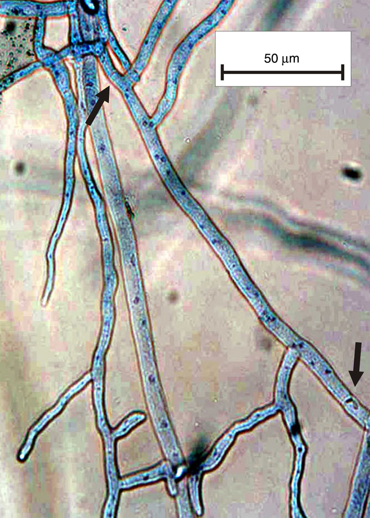
The genus Rhizoctonia DC (1805) has long been studied as an important soilborne pathogen that causes a wide variety of symptoms because it is a non-specialized pathogen3. Rhizoctonia sensu lato is characterized by the lack of conidiogenous cells and this taxon is composed of two groups based on the number of nuclei per cell: the multinucleate group that belongs to Rhizoctonia s. str. and the binucleate group that belongs to Ceratorhiza5. Currently, other authors consider the group a CeratobasidiumRhizoctonia complex7 and divide it into two groups: BNR (binucleate Rhizoctonia-like) and MNR (multinucleate Rhizoctonia-like)9. Many methods are used to observe the number of nuclei in fungal cells, e.g. safranine O, aniline blue, HCl-Giemsa. Some of these methods apply a staining solution involving laborious, time-consuming procedures that require no equipment (Fig. 1). Other methods use fluorophores, which are rapid and precise1,2,4,6,8,10.

Figure 1.

Multinucleate cells stained with lactophenol cotton blue. The arrows show the position of the septa.

(0,41MB).

Since a reliable and rapid method was needed to explore the MNR and BNR groups associated with rice crops in Argentina, we applied an accurate technique to observe the number of nuclei in the strain cells belonging to the CeratobasidiumRhizoctonia complex.

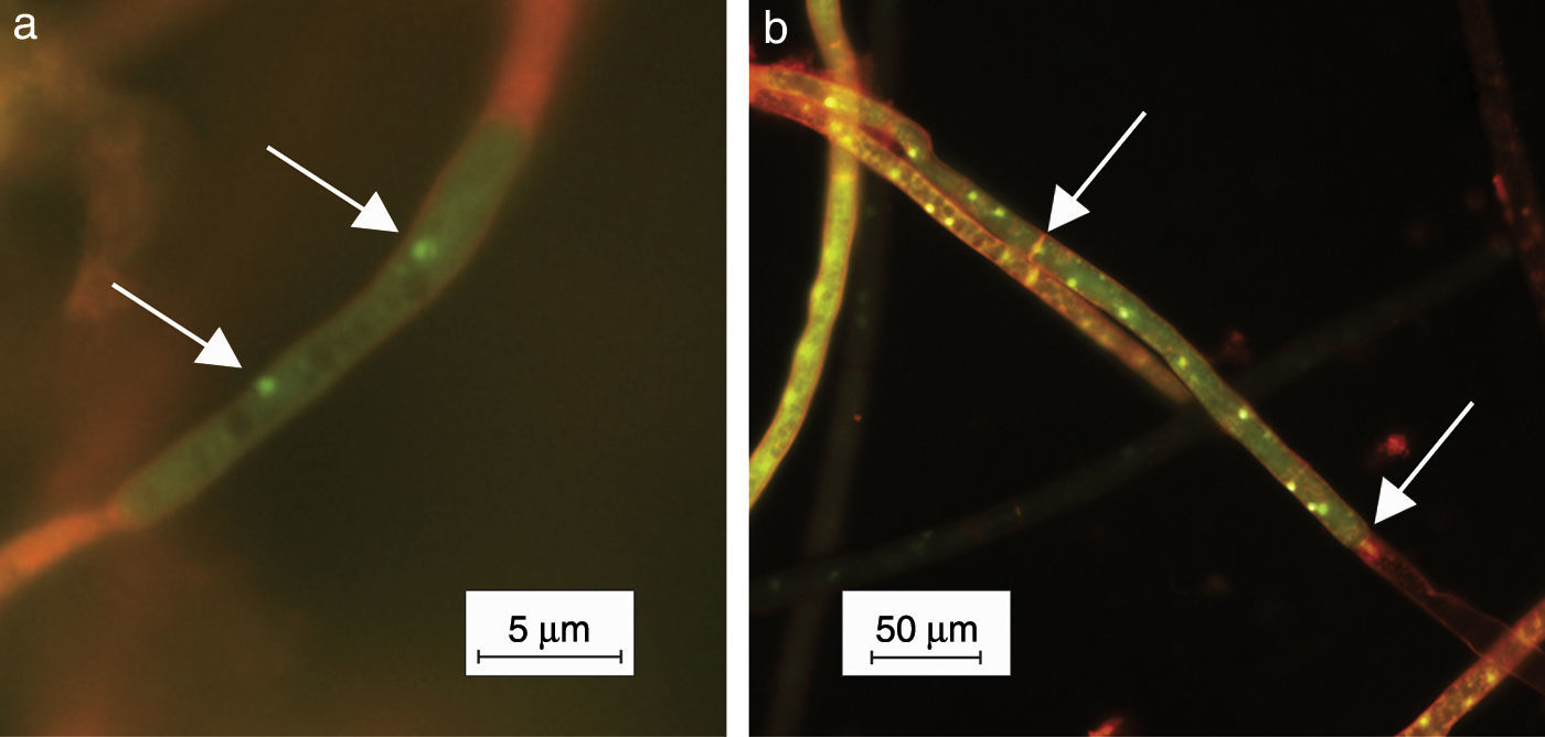
Small portions of mycelia grown on PDA for 24h were submerged in 0.01% acridine orange (Sigma–Aldrich, USA) aqueous solution during 10s. The method applied was a modification of the Yamamoto and Uchida's staining method11, the latter consisting of veronal buffer. The treated mycelium was observed under epifluorescence light using an OLYMPUS BX 51 microscope (Olympus, Japan). Digital photographs were taken using the Cool Snap-Pro System (Media Cybernetics, USA) (Fig. 2). The nuclei showed green fluorescence and the other cell components orange fluorescence. Thirty isolates from a collection of 36 were multinucleate and 6 were binucleate.

Figure 2.

Cells stained with acridine orange: (a) binucleate isolate; (b) multinucleate isolate. The arrows show the nuclei.

(0,09MB).

In comparison with the other methods mentioned above, the present method is characterized by easy handling and lower cost.

To our knowledge, this is the first time this methodology has been applied to observe nuclei in cells of Rhizoctonia isolates obtained from rice cultivars from Corrientes province, Argentina.

Ethical disclosuresProtection of human and animal subjects

The authors declare that no experiments were performed on humans or animals for this study.

Confidentiality of data

The authors declare that no patient data appear in this article.

Right to privacy and informed consent

The authors declare that no patient data appear in this article.

Funding, Financial support and acknowledgements

The present research was supported by the Governments of Argentina (Instituto Nacional de Tecnología Agropecuaria) and Japan (Japan International Cooperation Agency) in a bilateral collaboration program.

Conflict of interest

The authors declare that they have no conflicts of interest.

References
[1]
L. Cedeño, S. Mohali, E. Palacios-Prü.
Podredumbre radical causada por Rhizoctonia solani en mora de Castilla.
Fitopatol Venez, 6 (1994), pp. 34-37
[2]
L. Cedeño.
Método fácil y confiable para teñir núcleos en hongos del complejo Rhizoctonia.
Bioagro, 20 (2008), pp. 215-219
[3]
D. González, M. Cubeta, R. Vilgalys.
Phylogenetic utility of indels within ribosomal DNA and β-tubulin sequences from fungi in the Rhizoctonia solani species complex.
Mol Phylogenet Evol, 40 (2006), pp. 459-470
[4]
W.C. Kronland, M.E. Stanghellini.
Clean slide technique for the observation of anastomosis and nuclear condition of Rhizoctonia solani.
Phytopathology, 78 (1988), pp. 820-822
[5]
R.T. Moore.
The genera of Rhizoctonia-like fungi: Ascorhizoctonia, Ceratorhiza gen. nov., Epulorhiza gen. nov., Moniliopsis and Rhizoctonia.
Mycotaxon, 29 (1987), pp. 91-99
[6]
S. Oard, M.C. Rush, J.H. Oard.
Characterization of antimicrobial peptides against a US strain of the rice pathogen Rhizoctonia solani.
J Appl Microbiol, 97 (2004), pp. 169-180
[7]
F. Oberwinkler, K. Riess, R. Bauer, R. Kirschner, S. Garnica.
Taxonomic re-evaluation of the CeratobasidiumRhizoctonia complex and Rhizoctonia butinii, a new species attacking spruce.
Mycol Prog, 12 (2013), pp. 763-776
[8]
J.T. Otero, J.D. Ackerman, P. Bayman.
Diversity and host specificity of endophytic Rhizoctonia-like fungi from tropical orchids.
Am J Bot, 89 (2002), pp. 1852-1858
[9]
M. Sharon, S. Kuninaga, M. Hyakumachi, S. Naito, B. Sneh.
Classification of Rhizoctonia spp. using rDNA-ITS sequence analysis supports the genetic basis of the classical anastomosis grouping.
Mycoscience, 49 (2008), pp. 93-114
[10]
M. Sharon, B. Sneh, S. Kuninaga, M. Hyakumachi.
The advancing identification and classification of Rhizoctonia spp using molecular and biotechnological methods compared with the classical anastomosis grouping.
Mycoscience, 47 (2006), pp. 299-316
[11]
D.T. Yamamoto, J.Y. Uchida.
Rapid nuclear staining of Rhizoctonia solani and related fungi with acridine orange and with safranin.
Mycologia, 74 (1982), pp. 145-149
Copyright © 2014. Asociación Argentina de Microbiología
Opciones de artículo
Herramientas
es en pt

¿Es usted profesional sanitario apto para prescribir o dispensar medicamentos?

Are you a health professional able to prescribe or dispense drugs?

Você é um profissional de saúde habilitado a prescrever ou dispensar medicamentos

Quizás le interese:
10.1016/j.ram.2018.09.005
No mostrar más