


A 32-year-old woman from Venezuela presented to the Emergency department with epigastric pain, chills and fever during the last week. She reported no nausea, vomiting, jaundice or diarrhea. Physical examination revealed epigastric tenderness with no rebound. Blood tests showed AST 144U per liter (U/L) (reference range – RR-4–50), ALT 133U/L (RR 5–40), GGT 237U/L (RR 7–30), ALP 576 U/L (RR 42–141) and CRP 188mg per milliliter (RR<5) so an abdominal ultrasound was performed. It revealed a heterogeneous hepatic nodular lesion in the left liver lobe measuring 75×40×55mm. It was round and predominantly hypoechoic suggesting either a liver abscess or, less likely, a liver neoplasm. A CT scan was performed for better characterization, showing a low-density mass with a peripheral enhancing rim (Fig. 1). Blood and stool cultures were collected and empirical antibiotherapy to treat a liver abscess was started. Stool microscopy exam was negative but Entamoeba hystolitica IgG-ELISA was positive confirming an amebic liver abscess so a 10-day treatment with Metronidazole was completed. The patient did well and was discharged with Paromomycin for 1 more week to complete intestinal amebiasis treatment. The patient has been asymptomatic 8 months after discharge.
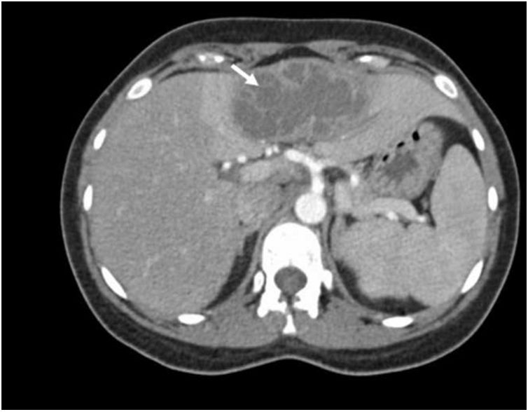
. It is possible to identify a central cavity filled with low-density fluid with internal septations. A peripheral enhancing rim corresponding to the abscess wall as well as a peripheral zone of surrounding oedema are seen. Amebic abscess are typically subcapsular single lesions located in the right liver lobe, although in this case is seen in the left lobe.")
Axial CT-scan showing a rounded, well-defined mass located in liver (arrow). It is possible to identify a central cavity filled with low-density fluid with internal septations. A peripheral enhancing rim corresponding to the abscess wall as well as a peripheral zone of surrounding oedema are seen. Amebic abscess are typically subcapsular single lesions located in the right liver lobe, although in this case is seen in the left lobe.
The authors declare no conflicts of interest.

