


The aim of this work was to determine the physico-chemical characteristics of the stained-glass windows into the 19th century of two mausoleums located in the city of Belém do Pará (Brazil), and to evaluate their state of conservation. The glass chemical composition was determined by WXRF and SEM/EDS. The samples’ morphology and the microorganisms’ identification were carried out by optical microscopy. The results indicated that the samples were soda-lime silicate glass, with approximately 70wt. % of SiO2, which contributed to the resistance of the stained glass to the weathering. The concentration of Na2O was normally twice the K2O, which contrasts with the composition of other panels produced during the same period, as reported in the literature. The biofilm is composed by cyanobacteria and rotifers. Overall, the panels analysed were in a good state of conservation, despite their exposure to tropical climate conditions for more than a century with no preventive measures whatsoever.
El objetivo del presente trabajo fue determinar las características físico-químicas de las vidrieras del siglo XIX correspondientes a dos mausoleos ubicados en la ciudad de Belém do Pará (Brasil) para evaluar su estado de conservación. La composición química del vidrio fue determinada por WXRF y SEM/EDS. La morfología de las muestras y la identificación de los microorganismos fueron realizadas por microscopia óptica. Los resultados indicaron que las muestras eran vidrios de silicato sódico-cálcico, con aproximadamente el 70% en peso de SiO2, lo que aumentó la resistencia a la corrosión de los vidrios de estas vidrieras. La concentración de Na2O fue normalmente el doble que de K2O, lo que contrasta con la composición de otros paneles producidos durante el mismo período, de acuerdo con la literatura. El biofilm presentó cianobacterias y rotíferas. En general, los paneles analizados presentaban un buen estado de conservación, a pesar de su exposición a las condiciones climáticas tropicales durante más de un siglo, sin las medidas de conservación preventivas.
Stained-glass windows are composed by coloured and colourless glass pieces fixed with lead structure. Usually, they are painted in the inner surface with a mixture of oxides and a bonding medium diluted with water, acetic acid or other solvents, which is called grisaille[1].
The decay of the stained-glass windows is influenced by different environmental parameters, such as temperature, humidity, air pollution, and microorganisms, individually or associated [2,3]. The main consequence is, the loss of transparency and the darkening of glass surface, which occur by the deposition of soot and dirt layers, and can induce the corrosion of the forming materials.
Different approaches have been taken concerning a variety of degradation factors, in stained-glass windows, mainly within European architectural heritage, among others, showing how complex the research in this field has become [4–7].
There are significant contributions regarding the chemical and physical composition of stained-glass windows from 19th century [8–12]. There are also works covering the characterisation of the other stained glass materials, such as the grisaille, the enamels [1,13,14] and the metallic support structure [15,16], which enables the better understanding of the panel as a whole.
The city of Belém, northern Brazil, has a great diversity of stained glass produced by different studios, which adorn historical buildings of a variety of architectural styles. The urban development of the city was considerably influenced by the Amazonia Rubber Boom of the late 19th century, when buildings began to incorporate window glass and stained glass, mostly commissioned from studios in Europe and south-eastern Brazil.
In general, these pieces have been exposed to temperatures above 24°C, a relative humidity around 55–90% and high rainfall [17], without preventive measurements. Given the lack of conservation, the original glasses of the windows with signs of deterioration were usually discarded. This non-desirable practice contributes to the gradual mischaracterisation of the architectural heritage of the city.
The aim of the present study is to investigate the conservation status of four stained-glass windows from 19th century mausoleums in the city of Belém. With this objective, the following specific goals were carried out: (a) to characterise the chemical composition of the samples; (b) to evaluate the morphology of stained-glass windows surface; (c) to identify the microorganisms existing on the biofilm deposited on colourless glass surfaces.
Materials and methodsSites descriptionThe stained-glass windows analysed in this paper belong to two mausoleums found in different cemeteries in the city of Belém do Pará, Brazil. One is the Nossa Senhora da Soledade cemetery, situated in the district Batista Campos. The last burial was in 1880, nevertheless the cemetery is still open for visits, due to its magnificent monuments. The second mausoleum is placed in the Santa Izabel cemetery, situated in the district Guamá and is still in use.
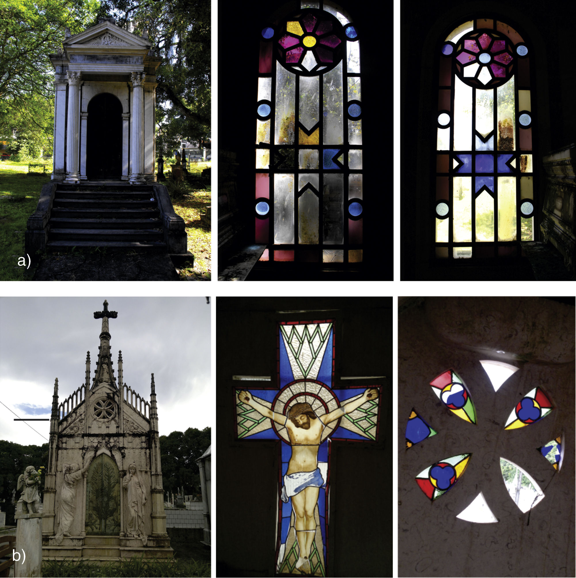
The mausoleum of the Assis Chermont family (ACM), located in the Nossa Senhora da Soledade cemetery, is the only monument in this place that has two stained-glass windows of coloured and colourless glasses. They were commissioned by Francisco Chermont and brought to Belém on the steamship Bernard & Bazil from Nantes (France) in 1881.
The building is situated in an area shaded by large trees (Fig. 1a). The articulated panels have a geometrical theme and are composed by colourless, blue, yellow, purple and opaque lilac pieces, with no grisaille or painting. It is also notable the existence of a thick green layer on the inner surface only on the colourless glass panes. Both panels appear to be in regular condition of conservation, except for a small gap in one window due to the fact that the mausoleum remains closed most of the time.
 Stained-glass windows in the Assis Chermont mausoleum in the Nossa Senhora da Soledade cemetery; (b) Stained-glass windows in the Britto Pontes mausoleum in the Santa Izabel cemetery; both in Belém do Pará (Brazil).")
The mausoleum of the family Britto Pontes (BPM), located in the Santa Izabel cemetery, has a stained glass and a rose window produced by the studio A Renascença (Lisbon, Portugal), probably from the end of the 19th century. The building is situated in the unshaded part of the cemetery. The rose window is in the main façade of the mausoleum and is composed by eight petals of red, blue, green and yellow glass, although three panes have now disappeared. A window with the form of a crucifix is installed in the opposite façade. It is composed of green, red and blue glasses, textured on the reverse surface, and colourless glasses painted with enamel on the obverse surface (Fig. 1b).
This panel presents considerably deterioration in comparison with the previous one, mainly due to a severe mechanical fatigue, which has damaged the panel's lead cames. Some of the glass panes have already been broken, while others have been displaced. In addition, some welds on the cames are deeply fractured. As a protective measure, a pane of corrugated glass was installed recently in the cruciform frame on the outside of the mausoleum.
It is important to note that none of the stained-glass windows described have been cleaned or restored in any way for more than a century. During this time, they have been exposed to an equatorial weathering. This is fundamental to understand the factors that have damaged the surface of the glasses selected for the present study.
Selection of the samplesTo characterise the 19th century stained-glass windows installed in the two mausoleums and to assess their current state of conservation, eleven samples of glass were selected: four from the Assis Chermont mausoleum (colourless, blue, opaque lilac, purple), and seven from the Britto Pontes mausoleum (green, yellow, blue, red flash, colourless with enamel, blue with texture). The panels were almost complete, with few gaps and displaced pieces. For this reason, the samples were selected based on the following criteria: (i) the colour of the glass, (ii) the accessibility, choosing preferentially damaged areas of the panels.
Characterisation techniquesThe samples were observed under optical microscopy (OM), and analysed by X-ray wavelength-dispersive fluorescence (WXRF) and scanning electron microscopy coupled to energy dispersive X-ray microanalysis (SEM/EDS). The optical microscopy was carried out using a Carl Zeiss Axiolab-Pol microscope (20× eyepiece and 10× objective lenses), attached to a G6 7.1 megapixel Canon camera to obtain images of the surface of the glass. Only the red flash glass sample was observed in cross-section.
The WXRF analyses were performed on powdered bulk samples using a PANalytical Axios Minerals WDS sequential spectrometer equipped with a ceramic X-ray tube, Rh-anode and 2.4 KW maximum power. The samples were prepared in two forms: (i) Fused disc for trace elements determination mixing 1g of sample and 6g of Li2B4O7 melt at 1000°C for 10min; (ii) Pressed powder samples for minor elements determination mixing 3g of sample and 0.9g of paraffin wax pressed at 20 tons’ load. The obtained data were processed using PANalytical SuperQ Manager software.
SEM/EDS analyses were carried out using a LEO-1430 VP. Images of samples’ surface and cross-section were acquired through the detection of secondary and backscattered electrons, with an attached energy dispersion system to produce semi-quantitative microanalyses, using acceleration voltages of 15kV and 20kV. Samples were coated with a conductive layer of Au (thickness=15nm) in an EMITECH K550X, at pressure of 2.10−1mbar, with current of 25mA for approximately ϕ2:30min.
Biological samplingSamples for the identification of microbial growth were obtained only from the Assis Chermont mausoleum. It was used a sterile swab previously wetted in distilled water, in order to allow the removal of all organic and inorganic matter, and then, carefully swabbed on the glass surface to collect the biofilm. Microorganisms were identified through optical microscopy.
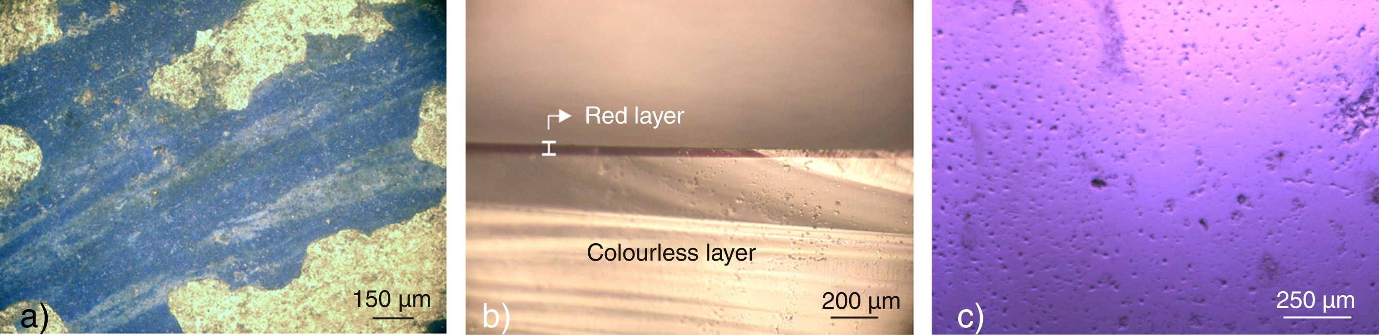
Results and discussionMorphology of the glass surface and alterationsThe optical microscopy revealed that in the sample BPM-6, the blue enamel has been detached from several points on the surface (Fig. 2a), what indicates that the vitrification temperature was too low for the adequate adhesion of the paint to the surface of the glass [1]. The flaking-off of the blue enamels from 17th to 20th centuries have been also attributed to the lack of flux in the painting and/or to the different thermal expansion coefficients between the enamel and the base glass [14,18,19].
 Detached painting in sample BPM-6; (b) Red layer of ∼40μm in the transverse section of the sample BPM-4; (c) Sample ACM-2, showing its rough surface.")
Sample BPM-4 is a flash glass with two layers of distinct colours. The thicker layer is colourless and the thinner one, with approximately 40μm, is red (Fig. 2b). The surface of sample ACM-2 was irregular with small pits (Fig. 2c), which may represent an initial phase of the corrosion process. The deposition of airborne particles of pollutants on the surface of the glass when it is wet, initiates a localised reaction between the particles and the network oxides of the glass [3,20], causing the visible deterioration of the material.
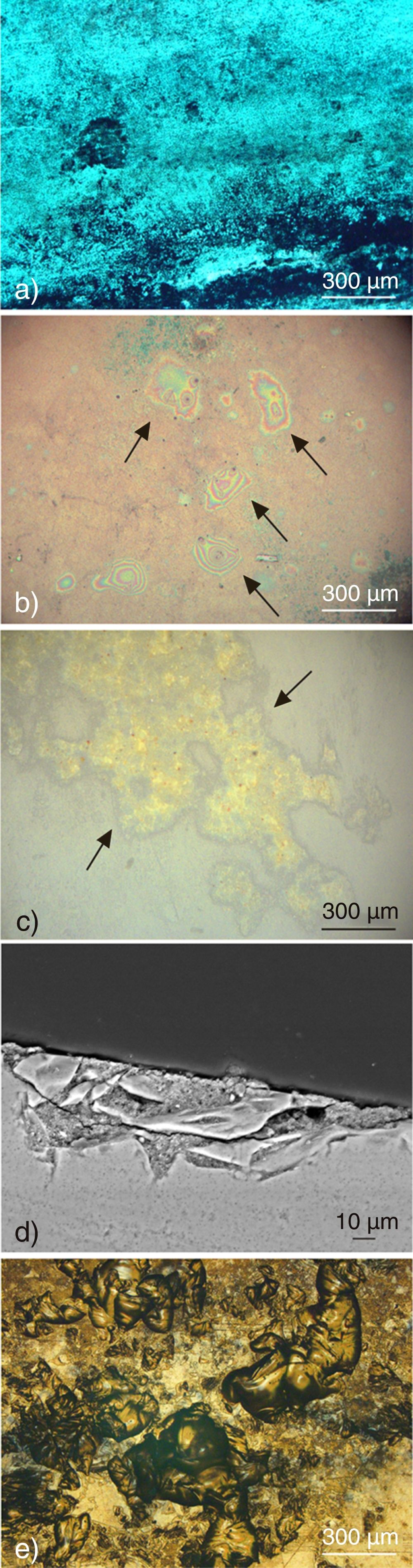
Regarding the alterations, a layer of dirt and soot was observed on the surface of all samples (Fig. 3a). The OM examination also revealed iridescent stains on sample ACM-4 (Fig. 3b), which can be related to the interaction of water and atmospheric pollutants on the glass surface [21]. Iridescence also indicates the formation of silica gel layers and it takes place when the glass is subjected to high relative humidity under stationary conditions, as it usually happens indoors, where condensation frequently occurs [22].
 Thick deposit of soot on the surface of sample BPM-1; (b) Visible iridescent stains on sample ACM-4; (c) Heterogeneous deposit in pits on the surface of sample ACM-3; (d) SEM image of sample ACM-3 in cross-section showing fractures on the glass in the area where the crust is formed; (e) Several interconnected pits on the surface of sample BPM-2.")
(a) Thick deposit of soot on the surface of sample BPM-1; (b) Visible iridescent stains on sample ACM-4; (c) Heterogeneous deposit in pits on the surface of sample ACM-3; (d) SEM image of sample ACM-3 in cross-section showing fractures on the glass in the area where the crust is formed; (e) Several interconnected pits on the surface of sample BPM-2.
The heterogeneous crust observed in sample ACM-3 (Fig. 3c) indicates that its distinct chemical composition may have favoured the corrosion process, which also induced fractures on the glass (Fig. 3d). Several interconnected pits, as seen on the surface of sample BPM-2 (Fig. 3e), were observed in all samples of coloured glass. These pits contribute to microbial growth and the accumulation of soot particles.
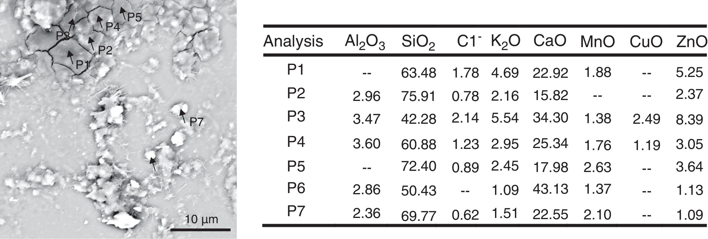
To better understand the occurrence of the iridescent stains, SEM/EDS analysis was performed on sample ACM-4. The results indicate that these stains represent the formation of a thick crust (Fig. 4). A semi-quantitative analysis of this area attested the presence of a large amount of Ca and C when compared to the amounts found on the glass surface, which may be attributed to the formation of a calcium carbonate (CaCO3) crust [7]. This fact can also represent the exposure of the matrix to microbial attack [23,24].
Chemical composition.")
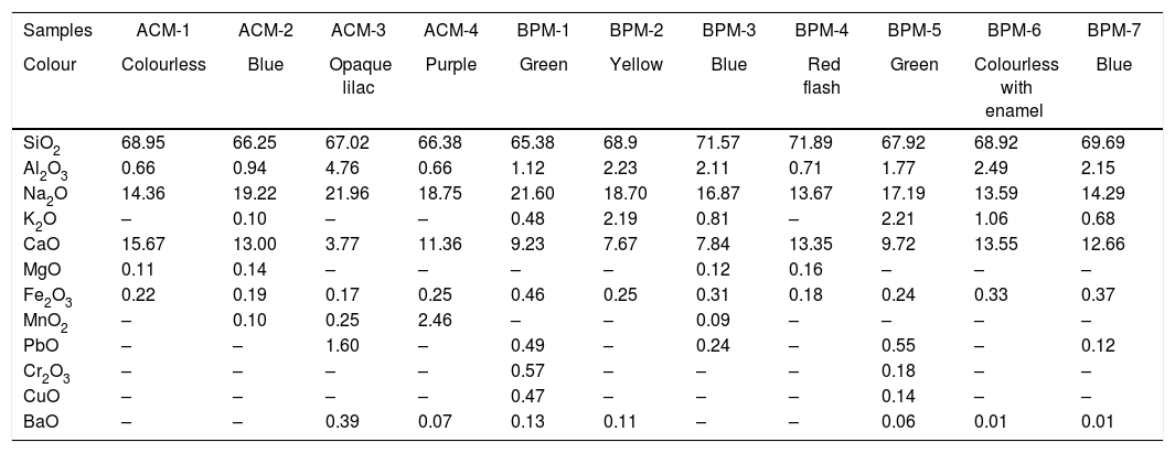
The WXRF analyses (Table 1) indicated that all the samples – both coloured and colourless – are soda-lime-silica glasses. The principal network-forming oxide is SiO2, which is around 70wt. % in all the samples. The network-modifying oxide is Na2O, with an average concentration of 17.29wt. %, and the network-stabiliser is the alkaline-earth CaO (10.71wt. %). Low concentrations of other oxides, such as the network-forming Al2O3 (1.78wt. %), the modifying K2O (1.08wt. %), and network-stabiliser MgO (0.13wt. %) were also detected, together with small amounts of transition metals such as Fe2O3 (0.27wt. %), MnO2 (0.73wt. %), and CuO (0.31wt. %), which could be related with the chromophores.
Chemical composition of the stained-glass windows, determined by wavelength-dispersive X-ray fluorescence in weight percent (wt.%) of oxides commonly used in glass.
| Samples | ACM-1 | ACM-2 | ACM-3 | ACM-4 | BPM-1 | BPM-2 | BPM-3 | BPM-4 | BPM-5 | BPM-6 | BPM-7 |
|---|---|---|---|---|---|---|---|---|---|---|---|
| Colour | Colourless | Blue | Opaque lilac | Purple | Green | Yellow | Blue | Red flash | Green | Colourless with enamel | Blue |
| SiO2 | 68.95 | 66.25 | 67.02 | 66.38 | 65.38 | 68.9 | 71.57 | 71.89 | 67.92 | 68.92 | 69.69 |
| Al2O3 | 0.66 | 0.94 | 4.76 | 0.66 | 1.12 | 2.23 | 2.11 | 0.71 | 1.77 | 2.49 | 2.15 |
| Na2O | 14.36 | 19.22 | 21.96 | 18.75 | 21.60 | 18.70 | 16.87 | 13.67 | 17.19 | 13.59 | 14.29 |
| K2O | – | 0.10 | – | – | 0.48 | 2.19 | 0.81 | – | 2.21 | 1.06 | 0.68 |
| CaO | 15.67 | 13.00 | 3.77 | 11.36 | 9.23 | 7.67 | 7.84 | 13.35 | 9.72 | 13.55 | 12.66 |
| MgO | 0.11 | 0.14 | – | – | – | – | 0.12 | 0.16 | – | – | – |
| Fe2O3 | 0.22 | 0.19 | 0.17 | 0.25 | 0.46 | 0.25 | 0.31 | 0.18 | 0.24 | 0.33 | 0.37 |
| MnO2 | – | 0.10 | 0.25 | 2.46 | – | – | 0.09 | – | – | – | – |
| PbO | – | – | 1.60 | – | 0.49 | – | 0.24 | – | 0.55 | – | 0.12 |
| Cr2O3 | – | – | – | – | 0.57 | – | – | – | 0.18 | – | – |
| CuO | – | – | – | – | 0.47 | – | – | – | 0.14 | – | – |
| BaO | – | – | 0.39 | 0.07 | 0.13 | 0.11 | – | – | 0.06 | 0.01 | 0.01 |
Obs.: (–) Below the detection limit.
The sample ACM-3, which is the opaque glass, presented a relatively high concentration of Al2O3 (4.76wt. %) and low of CaO (3.77wt. %). These concentrations are related to the techniques used to obtain the opaque effect of this glass [25]. The addition of aluminium compounds to the glass network increase their viscosity and create surface tensions, which confers greater resistance to thermal shock [25]. This high concentration may provide a different degree of protection against weathering.
The mean concentrations of Cr2O3 (0.66wt. %) and CuO (0.54wt. %) detected in two samples from the Britto Pontes mausoleum (BPM-1, BPM-5) were used as chromophores to obtain the green colour of the glass. On the other hand, the sample ACM-4 from Assis Chermont mausoleum present a high content of MnO2 (2.46wt. %), which was used to colour the glass in purple, whereas the chromophores in samples of blue glass (ACM-2, BPM-3, BPM-7) were below the limit of detection of the WXRF technique, as well as the following trace elements (0.01%): MnO, Zr, Sr, Cu, As, Pb, Rb, Nb, Co, Zn, V, Y, Cr, Ni, Ga and U.
Despite their different provenance, the stained-glass windows of both mausoleums have very similar chemical composition, considering the slight variation of the oxides amounts. Except for the MnO2, which is more frequent in Assis Chermont mausoleum, and PbO, which is more frequent in Britto Pontes samples.
The composition of the samples analysed here was also different from that of other 19th century stained-glass windows, investigated in previous studies [5,9,11]. The amount of SiO2 is higher (approximately in 6wt. %) for the studied samples. In contrast, the amount K2O for blue samples is lower (1.36wt. %), and the P2O5 is absent.
It is also interesting to note that the samples fall into two distinct groups, according to the Na2O:CaO ratio of the glass matrix. In the first group, which includes the samples ACM-1, BPM-4, BPM-6, and BPM-7 the amount of Na2O and CaO are similar (Fig. 5a). In the second group, which includes samples ACM-2, ACM-3, ACM-4, BPM-1, BPM-2, BPM-3, and BPM-5 the amount of Na2O is twice the CaO (Fig. 5b). The chemical composition of this second group is similar to the glass used in the modern restoration in St. Anthony's Basilica studied by Silvestri et al. [11], albeit with different colours.
 Group of samples corresponding to Na2O≈CaO ratio; (b) Group of samples corresponding to Na2O<CaO.")
Regarding the enamel applied on the surface of the sample BPM-6, EDS microanalysis revealed that both dark and light blue are mainly composed by CoO, ZnO and PbO, only varying the amounts (Fig. 6). However, as reported by Schalm et al. [14], it is more common to use Cu in light blue enamels, although Co, Fe and Mn can be also added in a small amount, what was not identified in the sample's painting. The very high concentrations of PbO are related to the flux employed on the paint [18].
Identification of microorganisms.")
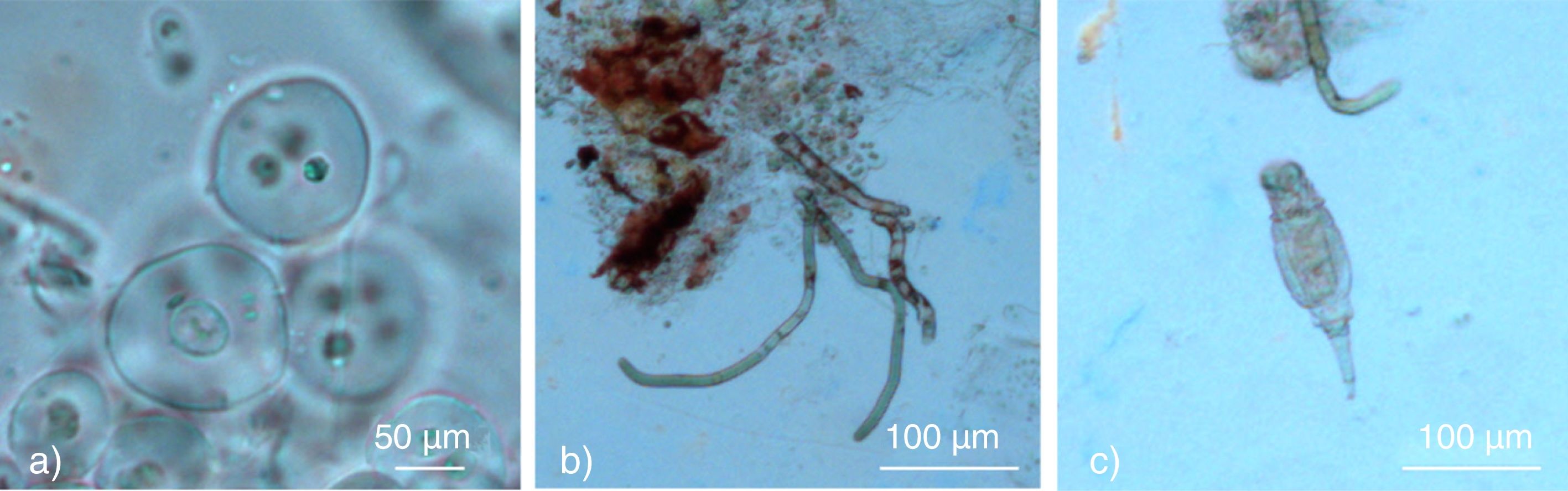
The biofilm found adhered only to the colourless glass panes of the Assis Chermont mausoleum was thick enough to allow the passage of sunlight through these panes. The OM examination suggested the presence of cyanobacteria of the genera Gloeocapsa and Oscillatoria, as well as rotifers of the genera Encentrum (Fig. 7). The last one is commonly found in freshwater environments composing the zooplankton community structure [26,27].
 Gloeocapsa sp.; (b) Oscillatoria sp.; (c) Encentrum sp. (Rotifers).")
These microorganisms play a crucial role in damaging the glass surface, excreting chemically aggressive substances (e.g. extracellular polysaccharides, polymers) and producing physical attack as well, since the glass network provides elements needed for their growth [6,28]. In the case of the rotifers, phosphorus and nitrogen compounds are released [29] and if the rates of these compounds are high, it creates the ideal environment to enhance the corrosion mechanisms on the glass surface. Additionally, phosphorus and nitrogen provide nutrient for algae, also creating a symbiosis between the different colonies within the microflora.
Nevertheless, the air condensation inside the mausoleum, caused by the fact of the windows have jammed shut, increases the growth of algae and cyanobacteria. Not to mention the great passage of sunlight through the colourless glass, which is essential for their development. This justifies the presence of biofilm only on the internal surface of the colourless glass panes, and never on the coloured glass. Moreover, it was also observed that the opacity created by the biofilm on the material surface, is more evident at the edges of the biofilm.
The presence of microorganisms in the local rainwater, such as diatoms (Navicula), can induce dark stains in the interface between the transparent glaze and the ceramic body of 19th century German tiles used to face historical buildings in Belém [30]. Given the similarities of glaze and glass, it is possible that the microorganisms of this genera also induce pitting on the surface of the glass, due to the fact that the silica is essential for their metabolism [31].
ConclusionsThe multi-analytical approach used in the present study, which included X-ray spectrometric techniques, proved to be useful for the characterisation of the French and Portuguese stained-glass windows installed in 19th century mausoleums in the Nossa Senhora da Soledade and Santa Izabel cemeteries, respectively. In addition, this study provides important data on the chemical composition of the glass, which contributed to the database on the conservation of stained-glass windows of this period under tropical climate conditions, which is still very poorly understood.
Despite being produced in different countries, the samples from the two mausoleums had the same general chemical composition, albeit two groups were distinct in basis with the relation of Na2O:CaO. The concentration of SiO2, K2O and P2O5 presented also considerable discrepancies. The glass samples analysed in this study were generally well conserved, mainly due to their high chemical stability. However, the polluted environment, combined with the prolonged exposure of the stained-glass windows to the humid equatorial climate typical of the northern Brazil could accelerate the glass deterioration.
Regarding the microbial community existing on the surface of the studied stained glasses, it is clear the symbiosis established between the cyanophyceae and the rotifers. The adherence of the biofilm only on the colourless glass panes validates the necessity of sunlight for the microorganisms’ growth, as well as the air condensation inside the mausoleum. The results of this study reinforce the need of the regular monitoring of the panels’ condition, considering precipitation levels and relative humidity, to guarantee the integrity of the panes, and ensure their historical and aesthetic quality in the context of the local architectural heritage.
Authors are grateful to the technical support of Scanning Electron Microscopy Laboratory (LABMEV, Universidade Federal do Pará), and Nucleus for Preservation and Restoration Technology (NTPR, Universidade Federal da Bahia). AMCP is indebted to Drs. Gisele Marques & Inês Coutinho for critical reading and improving the manuscript. This work has been partially financed by the CAPES through a PhD grant to AMCP (0418-14-5), the Brazilian National Council for Science and Technology (CNPq) through 484400/2011-8 and 552690/2011-2 projects and by the Ministério de Ciência e Tecnologia de Portugal through a post doctoral scholarship to TP (SFRH/BPD/108403/2015).
