Polychromatic corneal dystrophy is an unusual pre-Descemet dystrophy, about which there are very few publications. The findings are presented in a case series of four patients with polychromatic corneal dystrophy, using a slit lamp, specular biomicroscopy, and confocal microcospy.
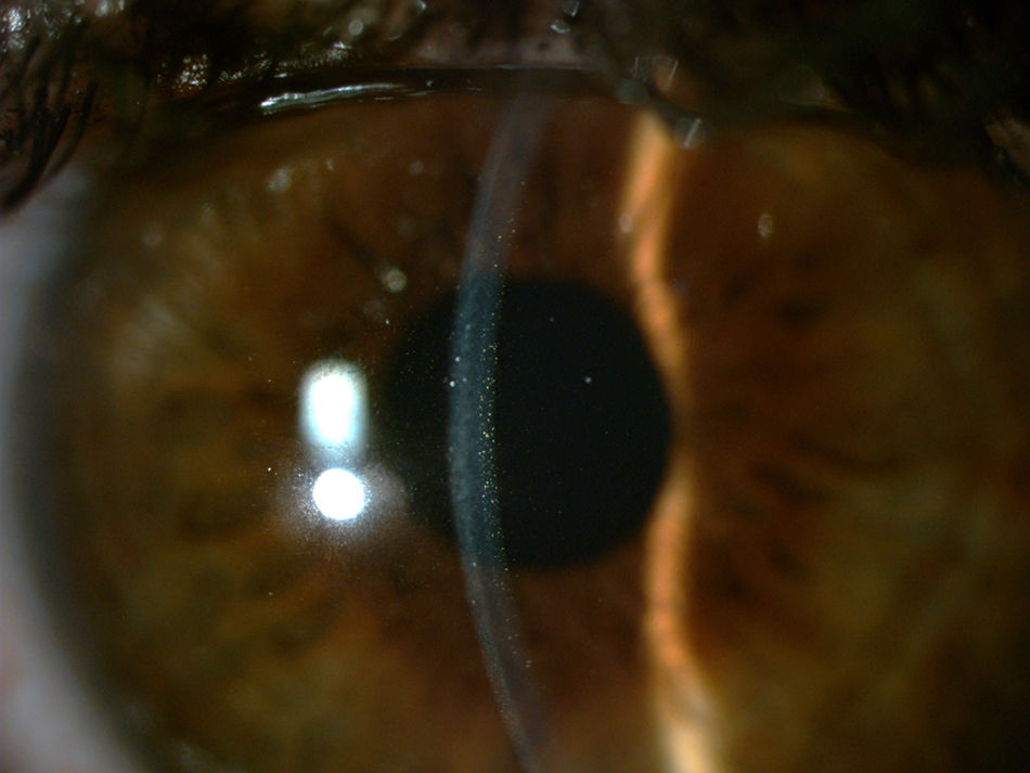
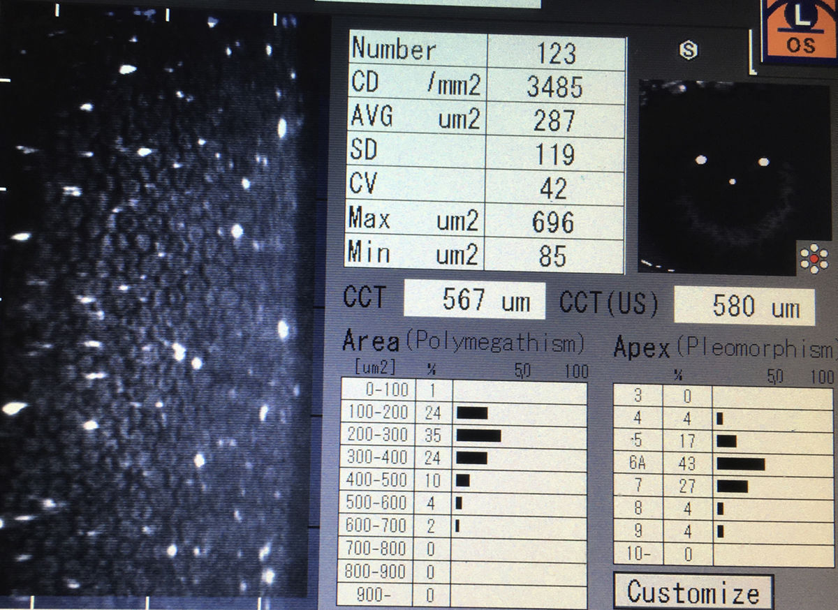
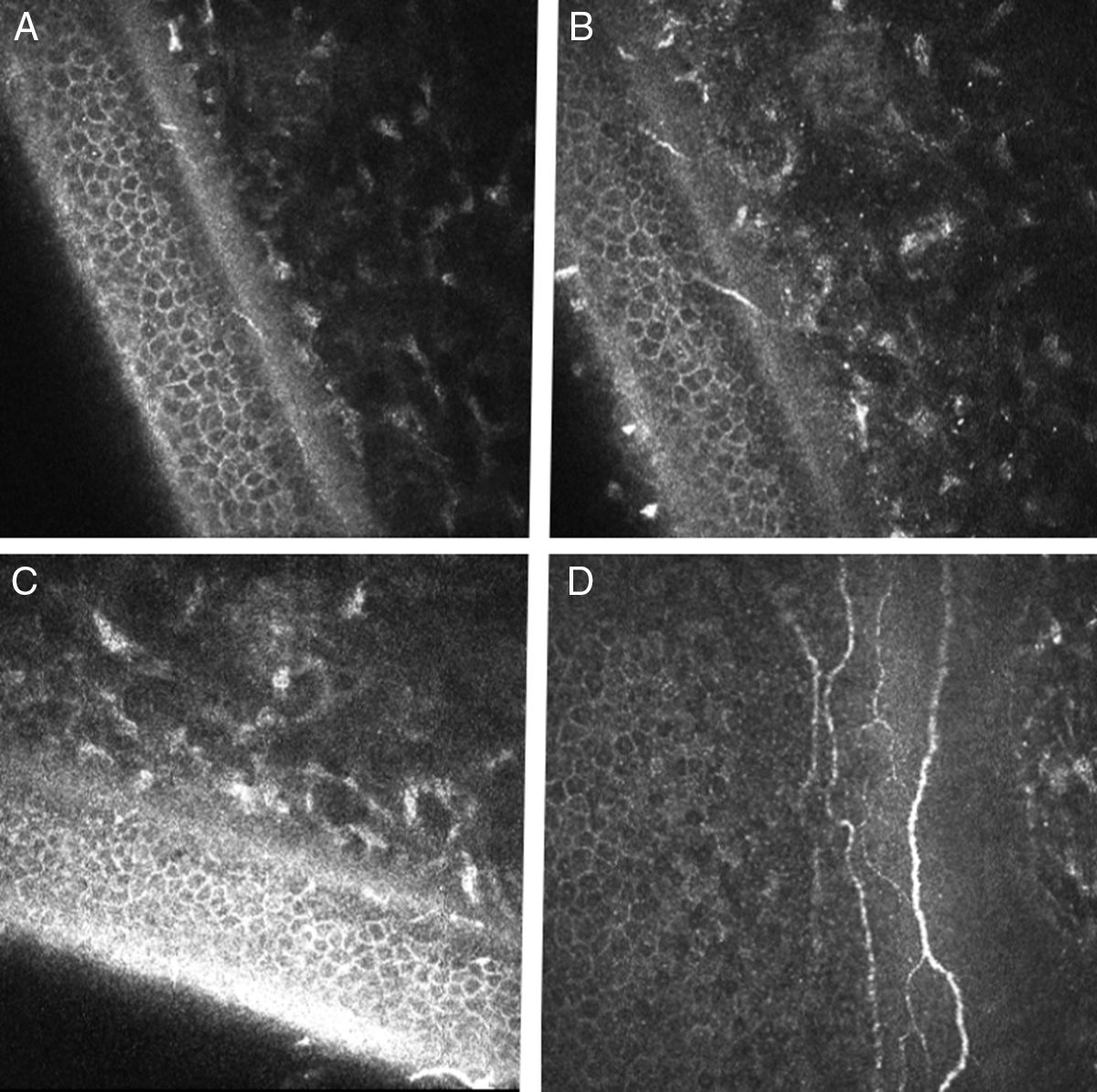
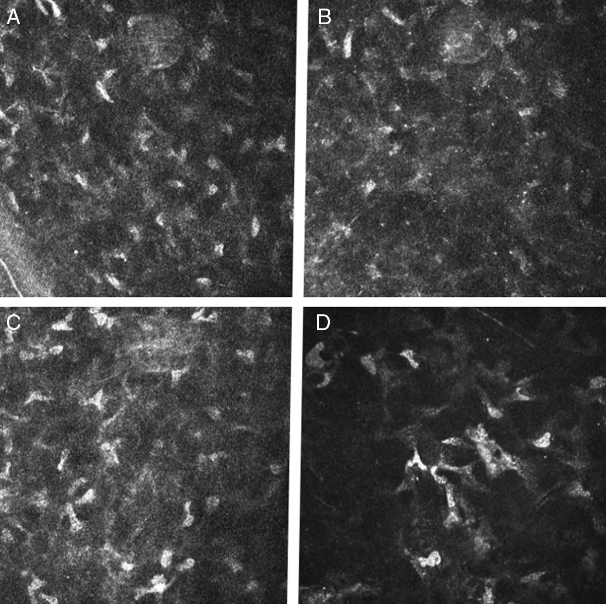
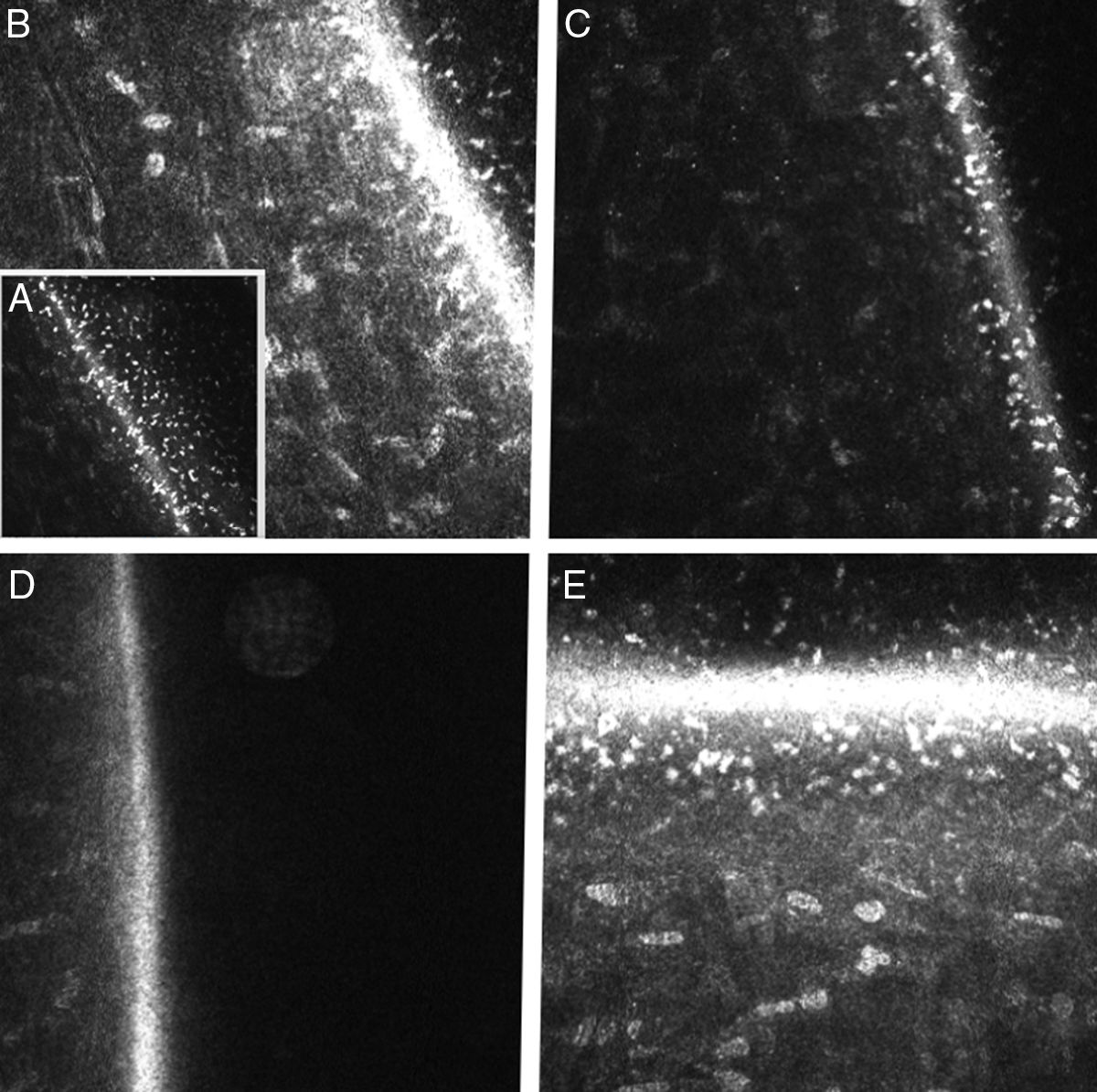
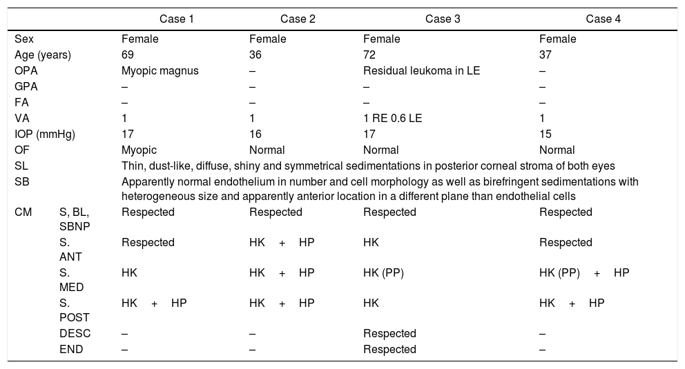
Clinical casesFour women, between 36 and 72 year-old, with the diagnosis of polychromatic corneal dystrophy in routine reviews. None reported visual symptoms or ocular history of interest. Anterior biomicroscopy showed multiple and small multicolored brilliant opacities in the posterior area of the corneal stroma, with normal epithelium and anterior stroma. The opacities were bilateral and distributed throughout the entire cornea. Direct family members were examined, but none of them showed opacities. In the specular biomicroscopy, a normal endothelium, with pre-Descemet hypereflective particles, was observed. With confocal microscopy, there were no abnormalities in epithelium, Bowman layer, or sub-basal nervous plexus. In two cases, the anterior stroma showed hyper-reflective keratocytes and with small hypereflective particles among them. In the middle stroma, hyper-reflective keratocytes were seen in the four cases, two of them showed tiny hypereflective particles, and in the other two there were abnormal keratocytes with prominent cytoplasmic processes. Posterior stroma in the four cases showed a lot of hypereflective keratocytes and hypereflective particles of different sizes. These particles prevented examining the endothelium.
ConclusionsPolychromatic corneal dystrophy has typical signs that allow it to be diagnosed and characterized. Although the biomicroscopy image only seems to show alterations in the posterior stroma, confocal microscopy shows that the dystrophy affects the entire corneal stroma.
La distrofia corneal policromática es una rara distrofia predescemética, con pocos casos publicados. Presentamos los hallazgos encontrados mediante lámpara de hendidura, biomicroscopia especular y confocal en una serie de 4 casos de esta entidad.
Casos clínicosSe trata de 4 mujeres de entre 36 y 72 años, diagnosticadas de distrofia corneal policromática en revisiones rutinarias. Ninguna refería síntomas visuales ni antecedentes oculares de interés. La biomicroscopia anterior objetivó múltiples y pequeñas opacidades brillantes multicolores en la zona posterior del estroma corneal, con respeto epitelial y estromal anterior. Las opacidades eran bilaterales y distribuidas por toda la córnea. Se exploró a familiares directos, los cuales no presentaban opacidades. En la biomicroscopia especular se observaba un endotelio normal con partículas hiperreflectivas predesceméticas. En la microscopia confocal no se objetivaron alteraciones en epitelio, capa de Bowman y plexo nervioso subbasal. En 2 casos se apreciaban en el estroma anterior queratocitos hiperreflectivos y pequeñas partículas hiperreflectivas entre ellos. En cuanto al estroma medio, aparecían queratocitos hiperreflectivos en los 4 casos, partículas hiperreflectivas de pequeño tamaño en 2 y en los otros 2 existían queratocitos anormales con procesos prominentes. El estroma posterior en los 4 casos mostró gran cantidad de queratocitos hiperreflectivos y de partículas hiperreflectivas de distintos tamaños. Tales partículas impedían la exploración del endotelio.
ConclusionesLa distrofia corneal policromática presenta signos típicos en la exploración, que permiten su diagnóstico y caracterización. Aunque la imagen biomicroscópica parece evidenciar alteraciones solo en el estroma posterior, la microscopia confocal demuestra que la distrofia afecta a todo el estroma corneal.