


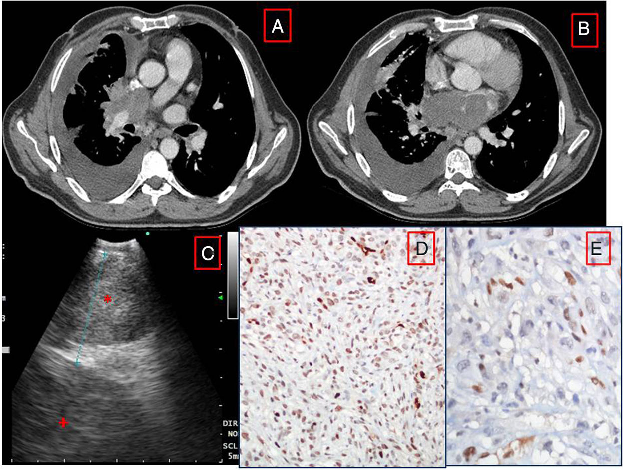
We report the case of a 50-year-old man who presented in an emergency department with a 3-day history of sore throat. A chest X-ray revealed right pleural effusion, which was confirmed by a CT scan of the chest. The chest CT also revealed that a mass originating from the left atrium (LA) was spreading to the hilar area (Fig. 1A and B). Transthoracic echocardiography confirmed that the lateral wall of the LA was thickened and that there was an inhomogeneous 48×32mm mass with an uneven surface in the LA, involving the mitral annulus and leaflets.
 CT scan showing the left atrial mass invading the right pulmonary artery. (C) EBUS image showing subcarinal area (*) and left atrial mass (+). (D and E). Histology of biopsy from the subcarinal area. The spindle cells are ERG(D) and Fli1(E) positive.")
The patient was referred for an EBUS, which revealed a subcarinal mass (Fig. 1C). A 21 G needle was used to aspirate the lesion, and the histology was consistent with cardiac angiosarcoma. The immunohistochemistry results were as follows: ERG(+), FLI-1(+), F8(+), vimentin(+), CD31(−), CD34(−), CD68(−), SMA(−) and Desmin(−) (Fig. 1D and E).
Malignant tumors of the heart are most often sarcoma (76–78%) and angiosarcoma, the most aggressive histologic type, accounts for about 31% of all primary cardiac malignant tumors, which usually originates in the right atrium; LA localization is rare.1 To the best of our knowledge, this is the first case of primary cardiac angiosarcoma diagnosed on an EBUS-guided sample.


