


To evaluate the contribution of 11C-Methionine PET in the early differentiation between tumor recurrence and radionecrosis in patients treated for a high grade glioma.
MethodThe study included 30 patients with glioma (III/IV grade) treated with surgery/radiotherapy/chemotherapy (5–8 months) and with an indeterminate MRI.
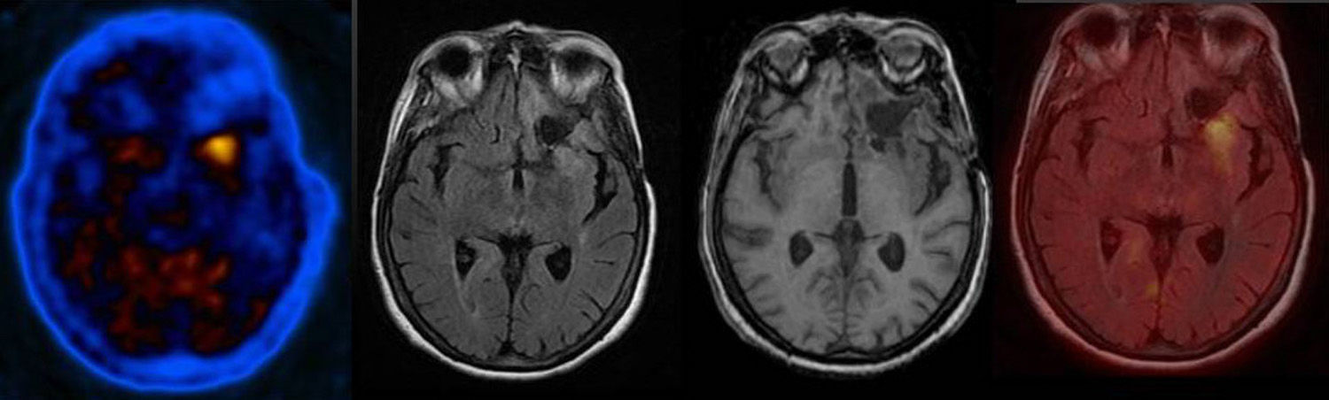
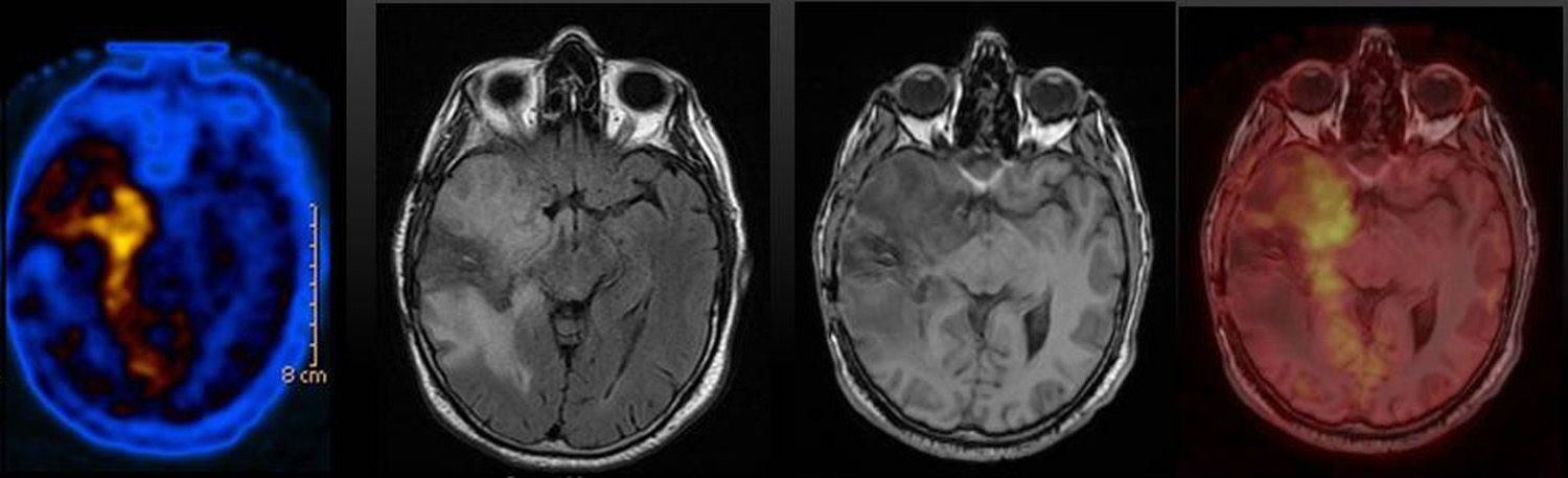
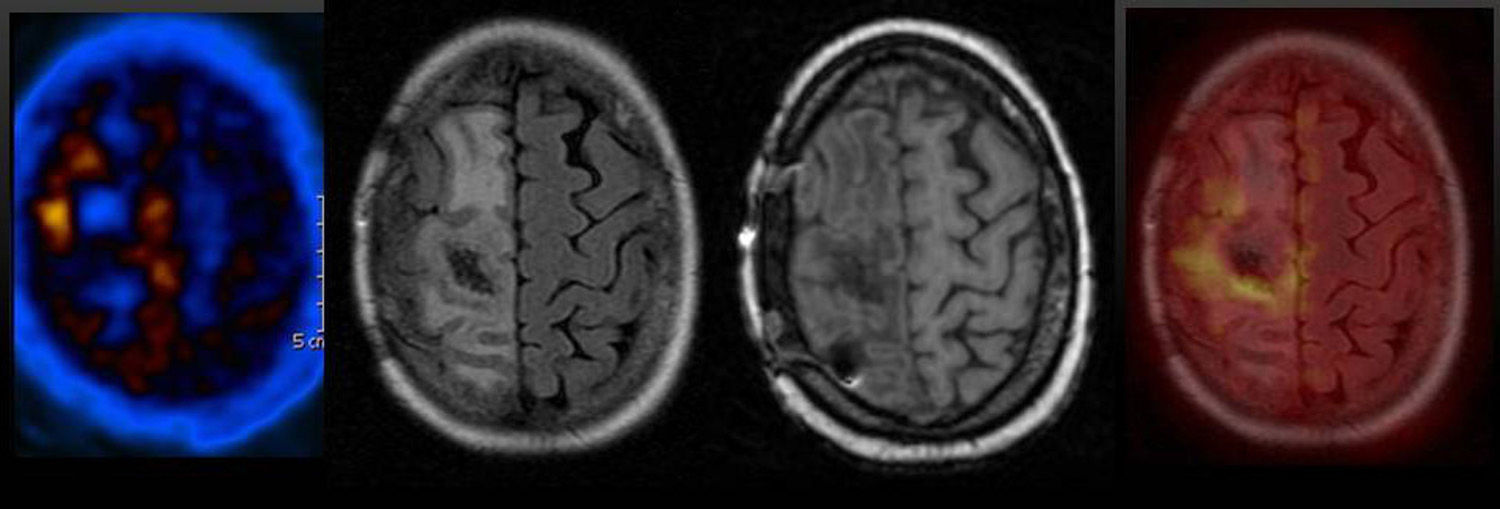
All patients underwent a 11C-Methione PET (within 15 days of MRI) and studies were visually analyzed (intensity and morphology of uptake), quantified (SUV max/SUV mean background), and coregistered to MRI (3D-Flair).
Patient management was decided by the neuro-oncology committee to clinical and imaging follow-up, second-line treatment, or surgery.
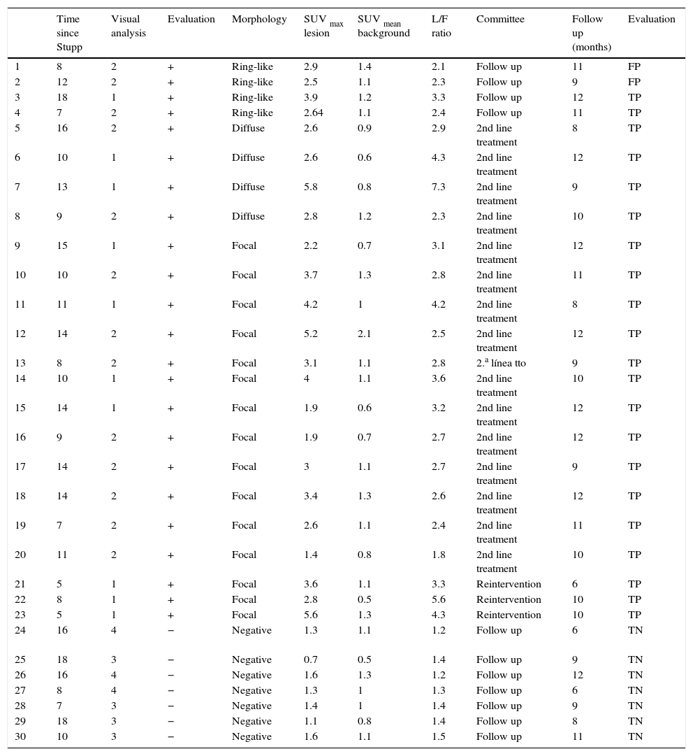
ResultsThere were 23 11C-Methionine PET studies visually positive. Morphology of uptake was focal in 15, diffuse in 4, and ring-shaped in 4.
Three out of the focal uptake cases underwent resection (Histopathology +). Sixteen underwent second-line therapy (11 responded; 5 progressed). The 4 cases with ring-shaped uptake were followed-up, and progression was found in 2 (true-positive), and disease-free in 2 (follow-up of 6 and 7 months, respectively) (false-positive).
Seven out of 11C-Methionine PET studies were visually negative, and all of them were disease-free (follow-up of 3–12 months).
SUV lesion/background was 2.79±1.35 in tumor recurrence, and 1.53±0.39 in radionecrosis (P<.05).
Taking into account a SUV lesion/background threshold of 2.35, the sensitivity and specificity values were 90.5% and 100%, respectively.
ConclusionVisual analysis, quantitative and PET/MRI coregistration of 11C-Methionine PET showed their complementary role in patients with indeterminate MRI results, thus allowing early differentiation between tumor recurrence and radionecrosis, and helping in the individual therapy approach.
Valorar la contribución de la PET con 11C-metionina en la diferenciación precoz entre recurrencia tumoral y radionecrosis en pacientes tratados de gliomas de alto grado.
MétodoTreinta pacientes tratados de glioma (grado III/IV) con cirugía/radioterapia/quimioterapia (5-18 meses) con RM indeterminada.
A todos se les realizó estudio de PET con 11C-metionina (<15 días tras RM) con análisis visual (grado de intensidad y morfología de captación), cuantificación (relación SUV máximo lesión/SUV medio fondo) y corregistro PET/RM (3D-Flair).
El manejo de los pacientes se decidió en el comité de neurooncología: seguimiento clínico-imagen, tratamiento de segunda línea o cirugía.
ResultadosVeintitrés estudios de PET con 11C-metionina fueron visualmente positivos. La morfología fue: 15 focales, 4 difusos y 4 anulares.
Tres de los focales fueron resecados (AP+). En 16 se realizó terapia de segunda línea (11 respuesta, 5 progresión). En los 4 de morfología anular se decidió seguimiento, con progresión en 2 (verdaderos positivos) y libres de enfermedad en 2 (6 y 7 meses después) (falsos positivos).
Siete estudios de PET con 11C-metionina fueron visualmente negativos, todos ellos libres de enfermedad (3-12 meses).
La relación SUV lesión/fondo en la recurrencia tumoral fue de 2,79±1,35 mientras que en la radionecrosis fue de 1,53±0,39 (p<0,05).
Con umbral de corte SUV lesión/fondo de 2,35 se obtuvo una sensibilidad y especificidad del 90,5 y 100%.
ConclusiónLa valoración de la PET con 11C-metionina, con análisis visual, cuantitativo y corregistro PET/RM muestra un papel complementario en los pacientes con RM no concluyente, permitiendo una diferenciación precoz entre recurrencia tumoral y radionecrosis, que ayuda a la individualización de la terapia.
Article
Revista Española de Medicina Nuclear e Imagen Molecular (English Edition)
