


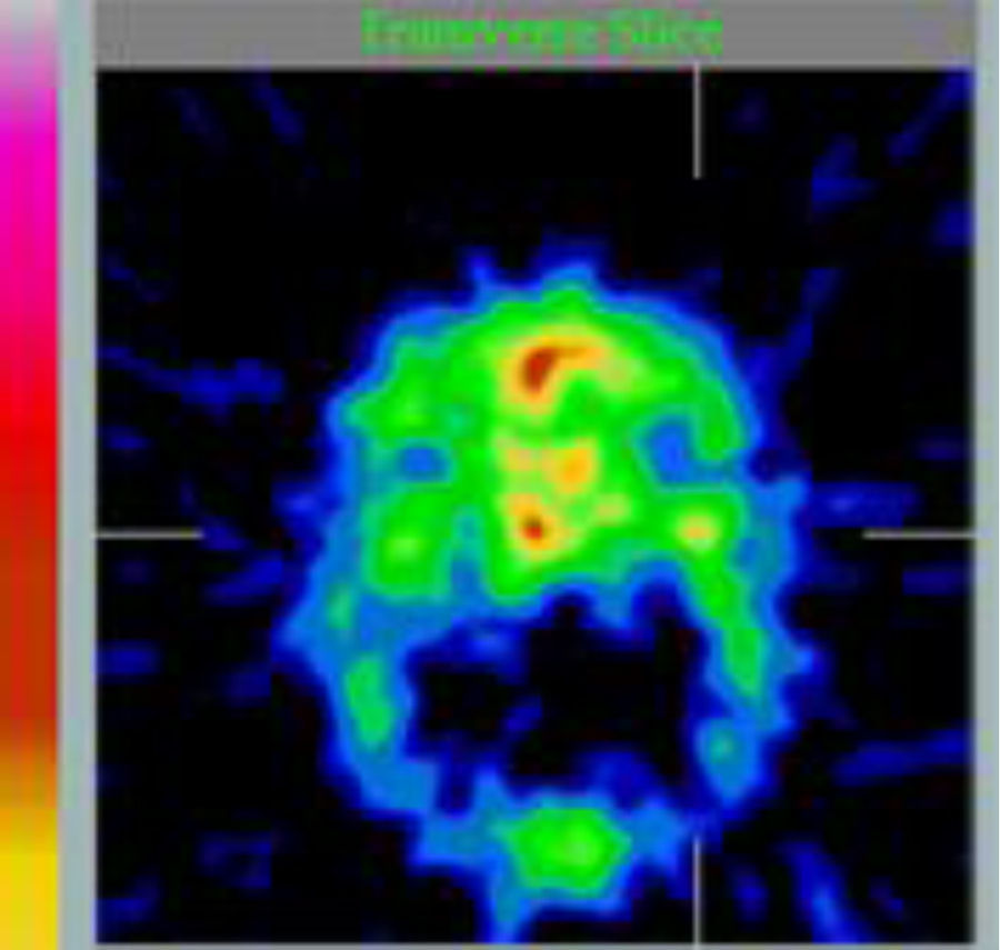
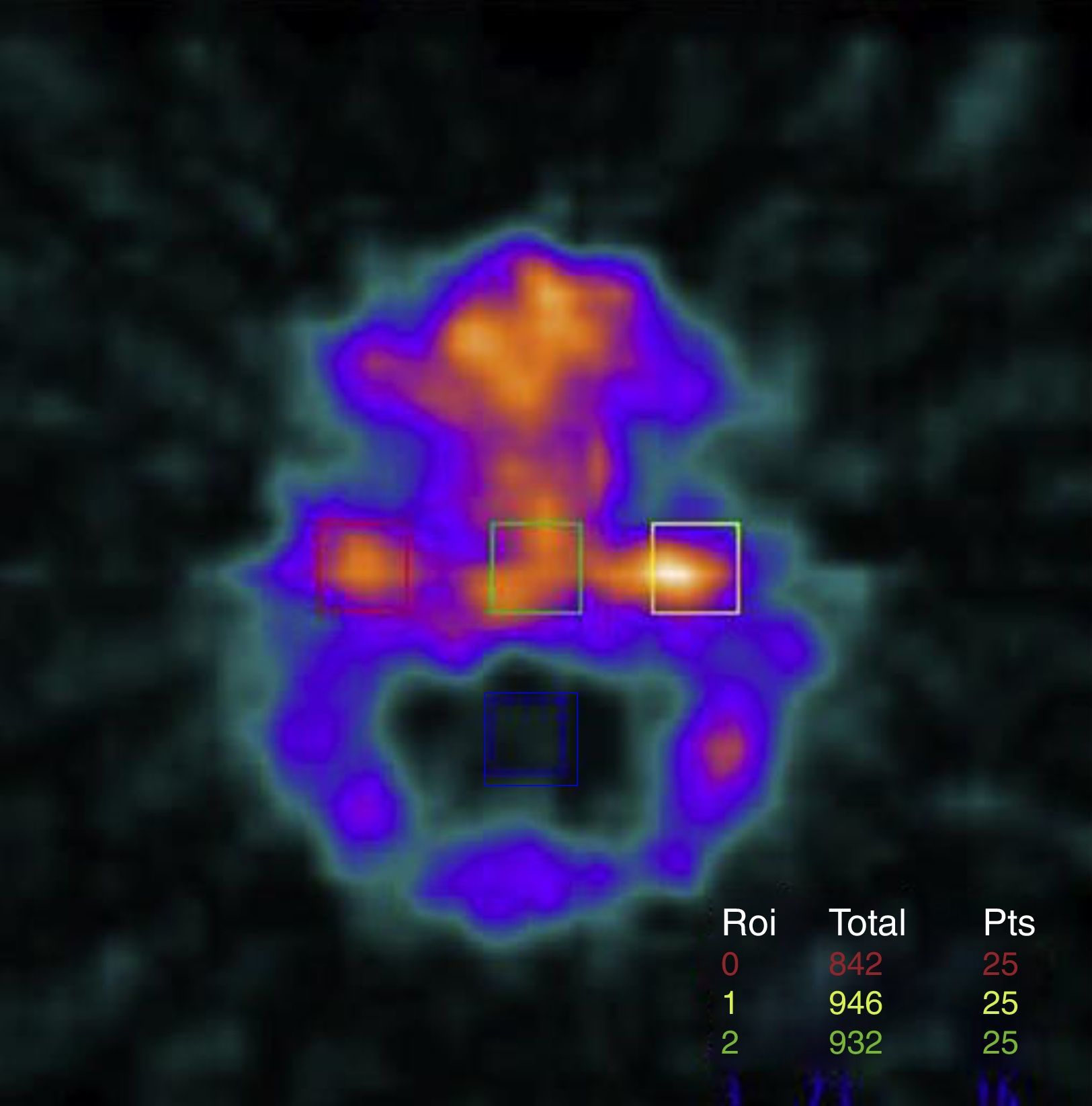
To compare qualitative vs quantitative results of Single Photon Emission Computerised Tomography (SPECT), calculated from percentage of 99mTc-MDP (methylene diphosphonate) uptake, in condyles of patients with a possible clinical diagnosis of condylar hyperplasia.
Materials and methodA retrospective, descriptive study was conducted on the 99mTc-MDP SPECT bone scintigraphy reports from 51 patients, with clinical impression of facial asymmetry related to condylar hyperplasia referred by their specialist in orthodontics or maxillofacial surgery, to a nuclear medicine department in order to take this type of test. Quantitative data from 99mTc-MDP condylar uptake of each were obtained and compared with qualitative image interpretation reported by a nuclear medicine expert.
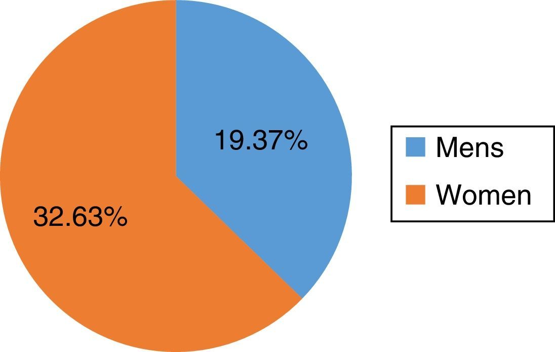
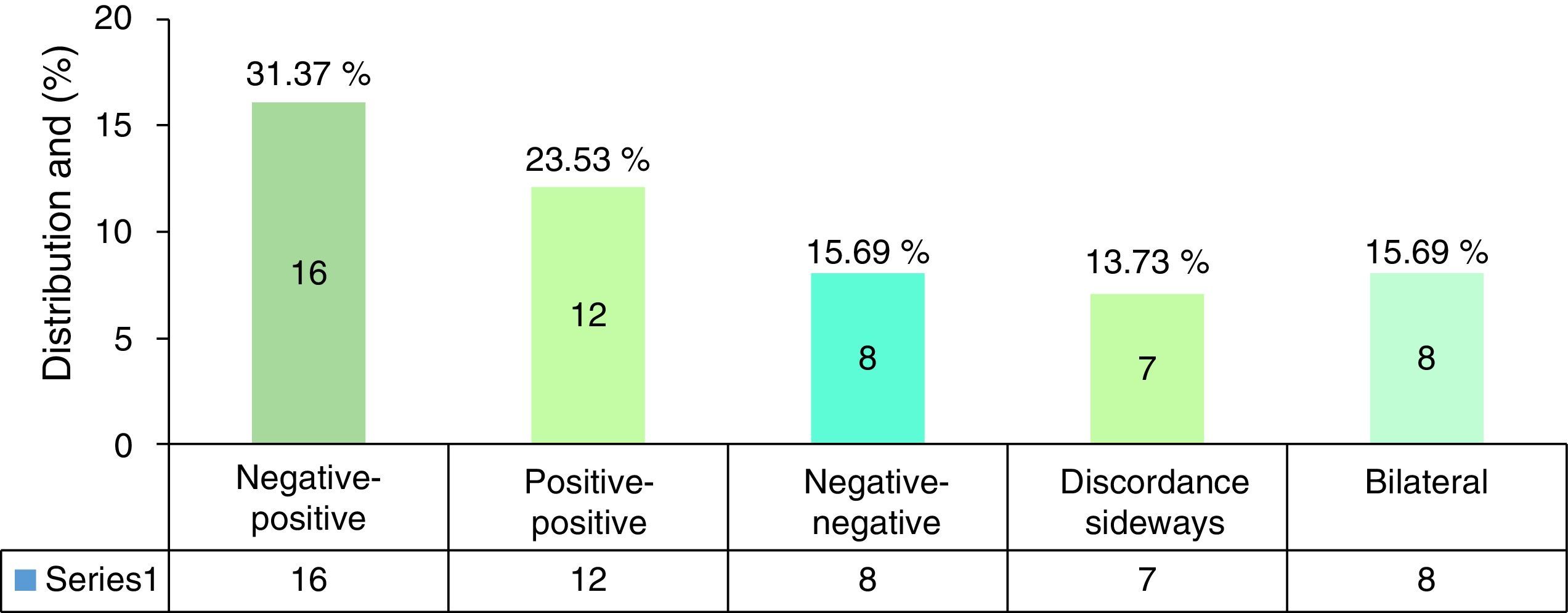
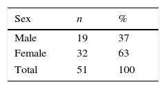
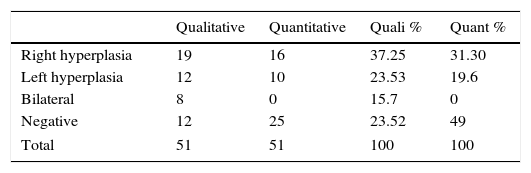
ResultsThe concordances between the 51 qualitative and quantitative reports results was established. The total sample included 32 women (63%) and 19 men (37%). The patient age range was 13–45 years (21±8 years). According to qualitative reports, 19 patients were positive for right side condylar hyperplasia, 12 for left side condylar hyperplasia, with 8 bilateral, and 12 negative. The quantitative reports diagnosed 16 positives for right side condylar hyperplasia, 10 for left side condylar hyperplasia, and 25 negatives.
ConclusionsNuclear medicine images are an important diagnostic tool, but the qualitative interpretation of the images is not as reliable as the quantitative calculation. The agreement between the two types of report is low (39.2%, Kappa=0.13; p>0.2).
The main limitation of quantitative reports is that they do not register bilateral condylar hyperplasia cases.
Comparar el resultado de los informes cualitativos de la tomografía computarizada de emisión por fotón único (SPECT), con los resultados cuantitativos, calculados a partir del porcentaje de captación del radiofármaco 99mTc-MDP (metilendifosfonato), en cóndilos de pacientes con sospecha clínica de hiperplasia condilar.
Materiales y métodoEstudio retrospectivo, descriptivo realizado en 51 pacientes con impresión clínica de asimetría facial y sospecha de hiperplasia condilar, remitidos a un centro de medicina nuclear para realizarles gammagrafía ósea-SPECT por el especialista en ortodoncia y/o cirugía maxilofacial. Se obtuvieron los datos cuantitativos del porcentaje de captación del radiofármaco 99mTc-MDP en ambos cóndilos, y se compararon con el informe cualitativo generado por el médico nuclear en cada uno de los sujetos.
ResultadosSe estableció la concordancia entre 51 informes cualitativos y sus resultados cuantitativos. Del total de la muestra, 32 eran mujeres (63%) y 19 hombres (37%). La edad de los pacientes estaba en un rango de 13-45 años (21±8 años). Según los informes cualitativos 19 pacientes fueron positivos para hiperplasia condilar derecha, 12 para izquierda, 8 bilaterales y 12 negativos. Según los resultados cuantitativos, 16 fueron positivos para hiperplasia condilar derecha, 10 izquierdos y 25 negativos.
ConclusionesLas imágenes de medicina nuclear son una importante herramienta diagnóstica, pero la interpretación cualitativa de la imagen no es tan confiable como la determinación cuantitativa. Los informes cualitativos concuerdan con los resultados cuantitativos de la prueba SPECT 99mTc-MDP, en un bajo porcentaje (39,2%, kappa=0,13; p>0,2).
La principal limitación del método cuantitativo es que no registra casos de hiperplasia condilar bilateral.
Article
Revista Española de Medicina Nuclear e Imagen Molecular (English Edition)
