Buscar en
Radiología (English Edition)
Toda la web
Inicio Radiología (English Edition) Diagnosing pneumothorax with ultrasonography
Journal Information
Vol. 56. Issue 3.
Pages 229-234 (May - June 2014)
Share
Share
Download PDF
More article options
Visits
2601
Vol. 56. Issue 3.
Pages 229-234 (May - June 2014)
Radiology through images
Diagnosing pneumothorax with ultrasonography
Diagnóstico ecográfico del neumotórax
Visits
2601
A. Lasarte Izcue
Corresponding author
aitorlasarte@gmail.com

Corresponding author.
, J.M. Navasa Melado, G. Blanco Rodríguez, I. Fidalgo González, J.A. Parra Blanco
Servicio de Radiodiagnóstico, Hospital Universitario Marqués de Valdecilla, Santander, Spain
This item has received
Article information
Statistics
Figures (6)
Show moreShow less
Tables (1)
Table 1. Comparison between the definition and the recommended therapy from the most important clinical guidelines on “large” pneumothoraces.
Additional material (5)
Abstract

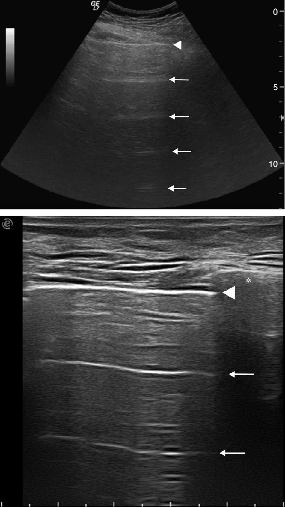
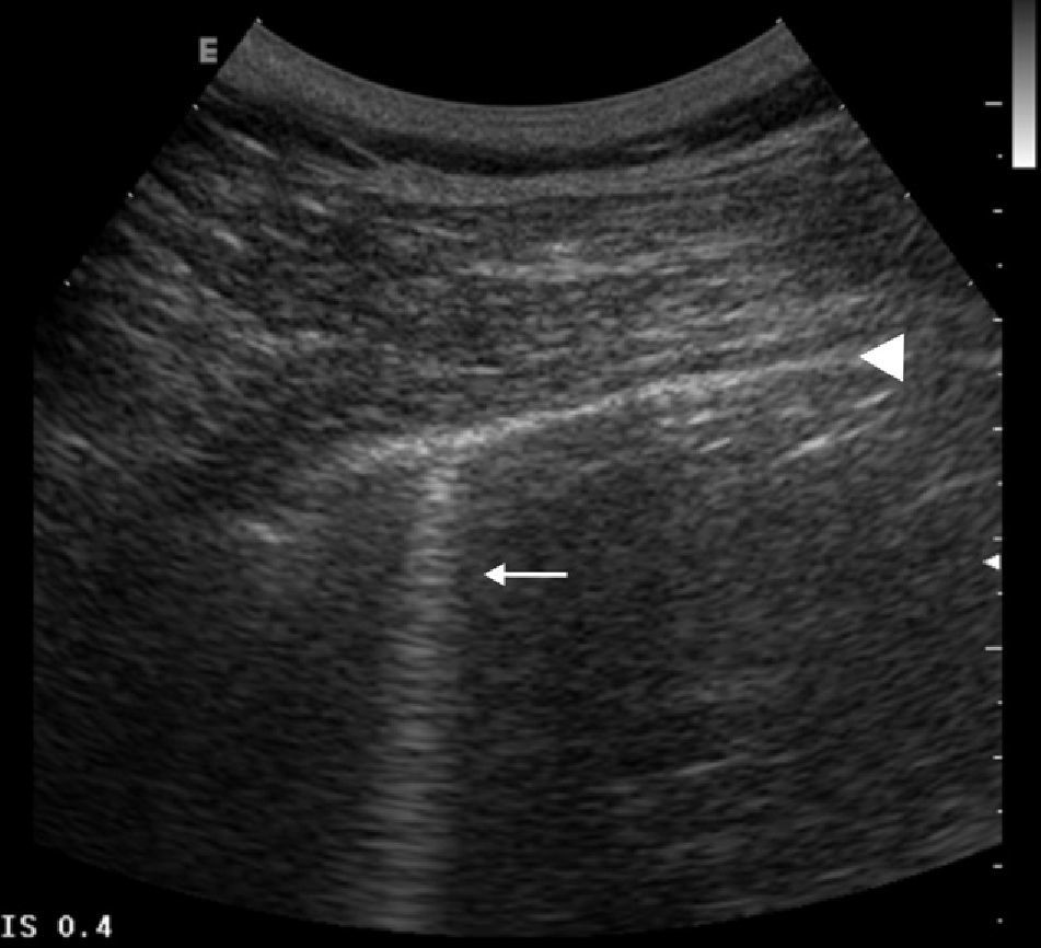
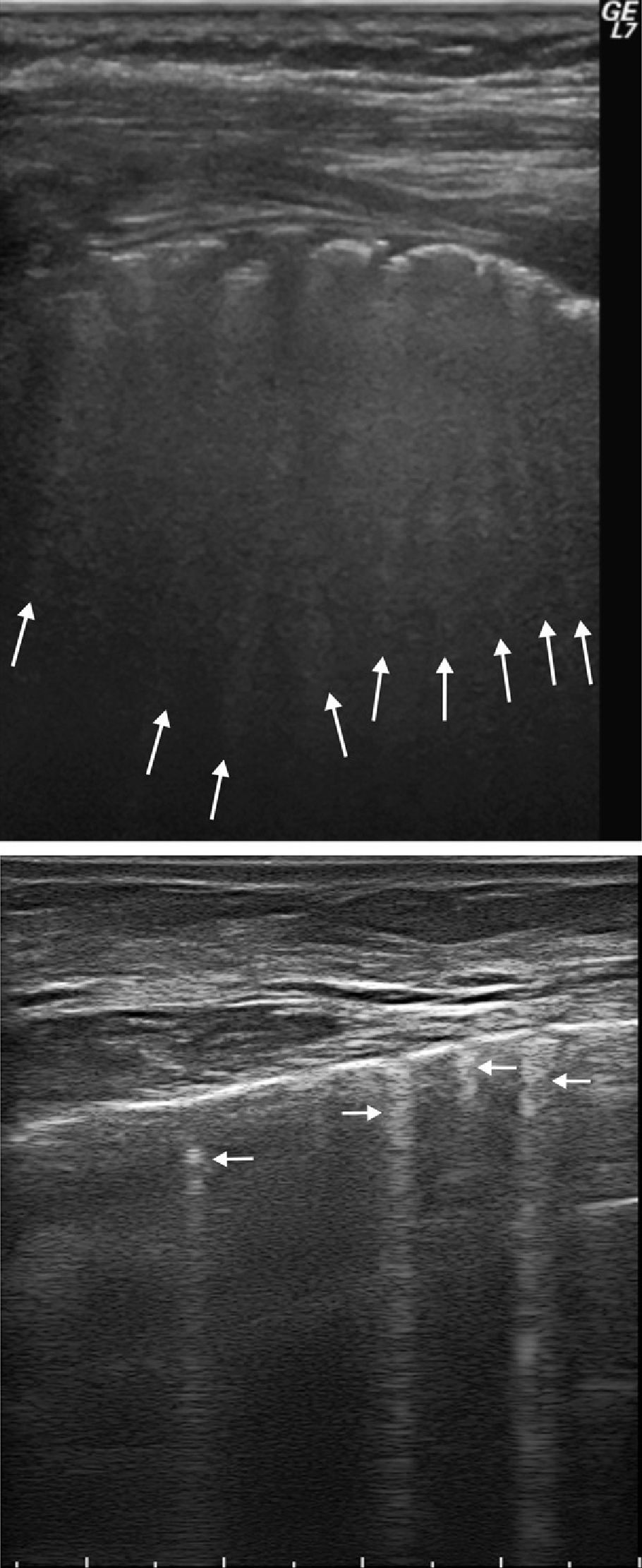
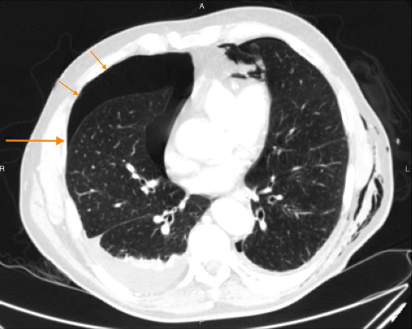
The ultrasonographic diagnosis of pneumothorax is based on the analysis of artifacts. It is possible to confirm or rule out pneumothorax by combining the following signs: lung sliding, the A and B lines, and the lung point. One fundamental advantage of lung ultrasonography is its easy access in any critical situation, especially in patients in the intensive care unit. For this reason, chest ultrasonography can be used as an alternative to plain-film X-rays and computed tomography in critical patients and in patients with normal plain films in whom pneumothorax is strongly suspected, as well as to evaluate the extent of the pneumothorax and monitor its evolution.

Keywords:
Pneumothorax
Lung
Chest
Ultrasonography
Diagnosis
Resumen

El diagnóstico ecográfico del neumotórax se basa en el análisis de artefactos. Combinando los siguientes signos: el deslizamiento pulmonar, las líneas A y B, y el punto pulmonar, es posible diagnosticar o descartar de forma segura la presencia de un neumotórax. Una ventaja fundamental de la ecografía pulmonar es su fácil acceso en cualquier situación crítica, especialmente en pacientes en la UCI. Por ello, la ecografía torácica podría utilizarse como alternativa a la radiografía simple y la TC en el paciente crítico, en pacientes con alta sospecha de neumotórax y radiografía normal, y para valorar la extensión del neumotórax y monitorizar su evolución.

Palabras clave:
Neumotórax
Pulmón
Tórax
Ecografía
Diagnóstico

Article

These are the options to access the full texts of the publication Radiología (English Edition)
Subscriber
Subscriber

If you already have your login data, please click here .

If you have forgotten your password you can you can recover it by clicking here and selecting the option “I have forgotten my password”
Purchase
Purchase article

Purchasing article the PDF version will be downloaded

Price 19.34 €

Purchase now
Contact
Phone for subscriptions and reporting of errors
From Monday to Friday from 9 a.m. to 6 p.m. (GMT + 1) except for the months of July and August which will be from 9 a.m. to 3 p.m.
Calls from Spain
932 415 960
Calls from outside Spain
+34 932 415 960
E-mail
Article options
Tools
Supplemental materials
es en pt

¿Es usted profesional sanitario apto para prescribir o dispensar medicamentos?

Are you a health professional able to prescribe or dispense drugs?

Você é um profissional de saúde habilitado a prescrever ou dispensar medicamentos