


A comprehensive study on deposition and conjugation of red emitting phosphor (GdVO4:Eu3+) and silver nanoprisms (Ag NP) on commercial single crystal silicon solar cell surface has been done to establish the optimum dielectric separating layer (PVA) sequence between consecutive species through confocal fluorescence mapping and spectroscopy. Results show that up to 310% fluorescence enhancement could be achieved in optimal arrangement of emitter, PVA and Ag NP layers on Si cell surface. The current work shows the potential of plasmon enhanced down shifting fluorescence of rare-earth doped vanadate in enhancing performance of SiPV devices.
Solar spectrum conversion through phosphors is a potential option for utilizing the ultraviolet (UV) and infrared (IR) part of the solar spectrum that otherwise remains unutilized by existing silicon solar cells. Silicon solar cells have inherent lower spectral response in the ultraviolet-blue (300–450nm) region because of its band gap (1.1eV) and fast surface recombination, high reflectance and thermalization losses of high energy photons (Shalav, Richards, Trupke, Krämer, & Güdel, 2005). Such losses may be minimized by using a down shifting (DS) phosphor layer which can convert a portion of the incident high energy UV photons to the visible region, where the Si solar cell has optimum spectral response (Chawla, Parvaz, Kumar, & Buch, 2013; Richards, 2006). Solar UV photons are absorbed near the Si cell surface which leads to the recombination of photo-generated electrons with surface defects, lowering the short wavelength spectral response. A DS phosphor layer on the Si surface can readily absorb UV photons and give fluorescence of lower energy photons that can be absorbed deep within the solar cell, close to the depletion region thus enhancing the collection efficiency. The red spectral region offers an optimum balance in the penetration depth and spectral response of Si cell. The development of suitable phosphors with high luminescence efficiency and integration of phosphor layer with Si solar cell is a daunting task. Rare earth-doped red-emitting phosphors, particularly Eu3+ doped vanadates (Gavrilović, Jovanović, Lojpur, & Dramićanin, 2014; Khan et al., 2008) and oxides (Dai, Foley, Breshike, Lita, & Strouse, 2011; Jayaramaiah, Lakshminarasappa, & Nagabhushana, 2012; Yadav et al., 2009) have shown very high luminescence efficiency. There are few reports on the direct deposition of phosphor layer on Si cell surface with dye (Klampaftis, Congiu, Robertson, & Richards, 2011), inorganic rare earth-doped phosphors (Yen-Chi, Woan-Yu, & Teng-Ming, 2011; Chen & Chen, 2011). Enhancement in Si cell efficiency by using Eu3+ complexes doped polyvinyl acetate (PVA) film (Liu et al., 2013; Le Donne, Acciarri, Narducci, Marchionna, & Binetti, 2009) and a layer of YVO4:Bi3+, Eu3+ nanophosphors (Huang et al., 2013) are reported. Recently there have been reports on the improvement in the performance of Si solar cells using nanostructured YVO4:Eu3+ downshifting phosphor layer (Chander et al., 2015; Kumar, Khan, & Chawla, 2013).
Gadolinium orthovanadate (GdVO4)-based materials have gained attention because of their interesting luminescent and magnetic properties. Gd compounds doped with luminescent lanthanide ions can be efficiently excited with UV radiation because of the strong absorption of the VO43− groups and efficient energy transfer from GdVO4 to lanthanide ions. Therefore, Eu3+, Dy3+, Sm3+ doped GdVO4 are widely used as phosphor (Singh et al., 2012).
Recent reports suggest that metal enhanced fluorescence is a viable option for further enhancement of rare earth (RE) emissions (Bishnoi, Das, & Chawla, 2014; Feng, Sun, & Yan, 2009) but optimal conjugation of phosphor and metal nanoparticles (MNP) is a challenge since surface plasmon resonance (SPR) of MNPs depend upon their shape, size and surrounding dielectric environment. However, contradictory reports exist in literature regarding efficiency enhancement of silicon solar cells by plasmonic effects including metal nanostructures (El Daif et al., 2012; Thouti, Sharma, Sardana, & Komarala, 2014; Wan et al., 2010) degrading the device's performance. Most reports on plasmon-enhanced fluorescence are on glass/quartz substrate (Buch, Kumar, Mamgain, & Chawla, 2013). A dielectric intervening layer between phosphor and MNPs play a crucial role in enhancing or quenching (Anger, Bharadwaj, & Novotny, 2006; Wang et al., 2011a; Wang et al., 2011b) fluorescence from phosphor material. Since optimum conjugation of substrate, phosphor and MNPs are crucial to perform plasmon enhanced fluorescence, it is extremely important to evolve the conjugation sequence on the Si solar cell surface. We have chosen an intense red-emitting phosphor GdVO4:Eu3+ with excitation range in the UV-blue region. Ag nanoprisms are chosen since they can strongly confine and enhance the EM field in the visible region because of their ability to exhibit distinct dipolar and quadrupolar resonances (Hao & Schatz, 2004) which can be tuned in the UV-visible region that can play an important role in fluorescence enhancement of RE ions (Wang et al., 2011a; Wang et al., 2011b). Polyvinyl alcohol (PVA) has been chosen as the spacer layer as it has been reported that a PVA layer on the Si cell surface does not change the cell's performance (Liu et al., 2013) and Eu3+-doped PVA layer on the Si cell has been shown to improve external quantum efficiency in the UV spectral region (Guedje, Giloan, Potara, Hounkonnou, & Astilean, 2012). We have done a systematic study of conjugating GdVO4:Eu3+ phosphor and silver nanoprisms so that maximum fluorescence enhancement could be realized on commercial Si solar cell surface.
2Experimental methodsGdVO4:Eu3+ was synthesized using a controlled solid state reaction method. For the synthesis of Eu (5%)-doped GdVO4 particles, analytical grade commercial Gd2O3 (99.99% pure), V2O5 and Eu2O3 (99.99% pure) have been used. The precursor materials were taken in their stoichiometric ratios, mixed thoroughly, packed in recrystallized alumina boat and fired at 1000°C for 2h in air atmosphere. The resulting product was allowed to cool slowly at room temperature and was crushed to a fine powder using mortar and pestle.
A silver nanoparticle solution was prepared by following the method by Heredia (2011). The silver precursor used was AgNO3 in which poly vinyl pyrrolidone was added followed by trisodium citrate (Na3C6H5O7) with continuous stirring. A solution of NaBH4 was added for reduction of silver salt to Ag+ followed by the formation of spherical Ag nanoparticles which were etched with H2O2 solution (30%w/v) to form silver nanoprisms.
A paste of phosphor GdVO4:Eu3+ with the consistency of paint was prepared in an appropriate amount (1g) of polymethyl methacrylate (PMMA) dissolved in 5ml dichloro methane. A dilute solution (3%) of polyvinyl alcohol (PVA) was prepared in DI water to be used as an intervening dielectric layer. The sequence of deposition of phosphor paint on single crystal Si solar cell (with lab tested efficiency of 13.6%), Ag NP colloidal solution and PVA solution on Si solar cell surface were varied in four different ways to optimize maximum fluorescence enhancement. The sequences adopted were (1) Si cell+GdVO4:Eu3++AgNP; (2) Si cell+GdVO4:Eu3++PVA+Ag NP; (3) Si Cell+PVA+GdVO4:Eu3++Ag NP; (4) Si cell+PVA+GdVO4:Eu3++PVA+Ag NP. Controlled drop casting of individual species followed by drying was harnessed for sequential deposition. The dielectric PVA layer was used to separate different species and to investigate the effect of an intervening dielectric layer on the fluorescence output from GdVO4:Eu3+.
2.1CharacterizationThe X-ray powder diffraction (XRD) of the Eu doped GdVO4 powdered sample was carried out on a Rigaku miniflex X-ray diffractometer with Cu-Kα radiation (1.54Å). The morphology of the powdered sample was inspected using a LEO 440 PC digital scanning electron microscope (SEM). The absorption spectra of the Ag NP solution was measured using Avantes UV-visible spectrometer. The photoluminescence (PL) characteristics were studied under a WITec Confocal fluorescence microscope (WITec 300M+) with PL mapping facility. Confocal fluorescence mapping and spectroscopy of the identical area was done of the bare Si cell and after each stage of deposition of GdVO4:Eu3+ and Ag NP solution and also PVA in samples (2–4) as mentioned above. A specific area of the cell was chosen under a 20× objective in the confocal microscope. Starting from the bare Si cell, confocal measurements were done for all 4 sets of layered structures and fluorescence distribution was recorded under excitation of a UV diode laser (λ∼375nm, output power 10mW, Toptica). Confocal fluorescence mapping and spectroscopy was done on the Si cell with sequence of layers deposited on it. In order to investigate the plasmonic effect on the red emission from Eu3+ doped in GdVO4 particles with various layer sequence of intervening PVA layer, the same area of the Si cell was used in each set, without disturbing the Si cell position on the confocal microscope sample plate. Spacer layer of PVA, phosphor layer and Ag NP layer was deposited on the Si cell surface by drop casting keeping the Si cell position fixed.
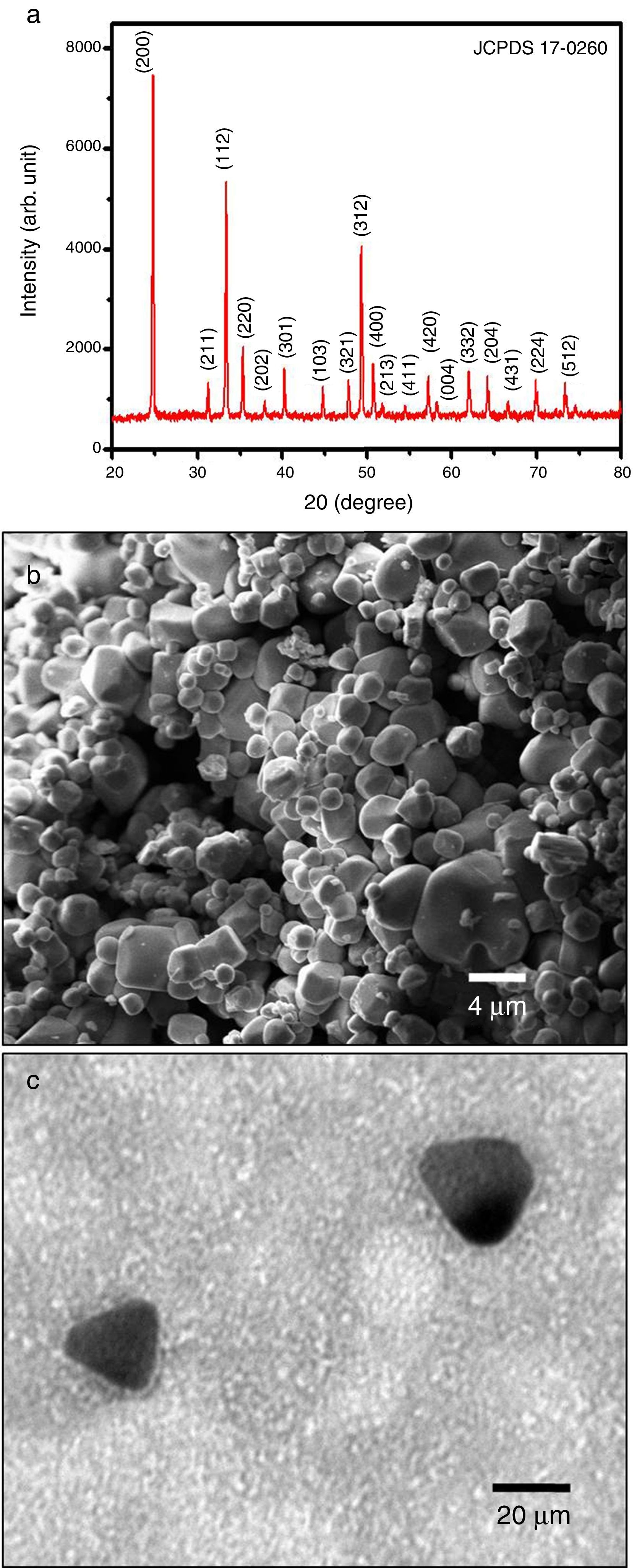
3Results and discussion3.1XRD, SEM AND TEMThe synthesized GdVO4:Eu3+ was found to be well crystalline with tetragonal phase (JCPDS No. 17-0260) as shown in Figure 1a. The SEM micrograph (Fig. 1b) shows GdVO4 particles with distinct rounded morphology. The HRTEM image of synthesized Ag nanoparticles displays that Ag nano prisms of average edge length 22nm are formed as shown in Figure 1c.
 X-ray powder diffraction spectrum of GdVO4:Eu3+; (b) SEM image of GdVO4:Eu3+; (c) TEM image of Ag nanoprisms.")
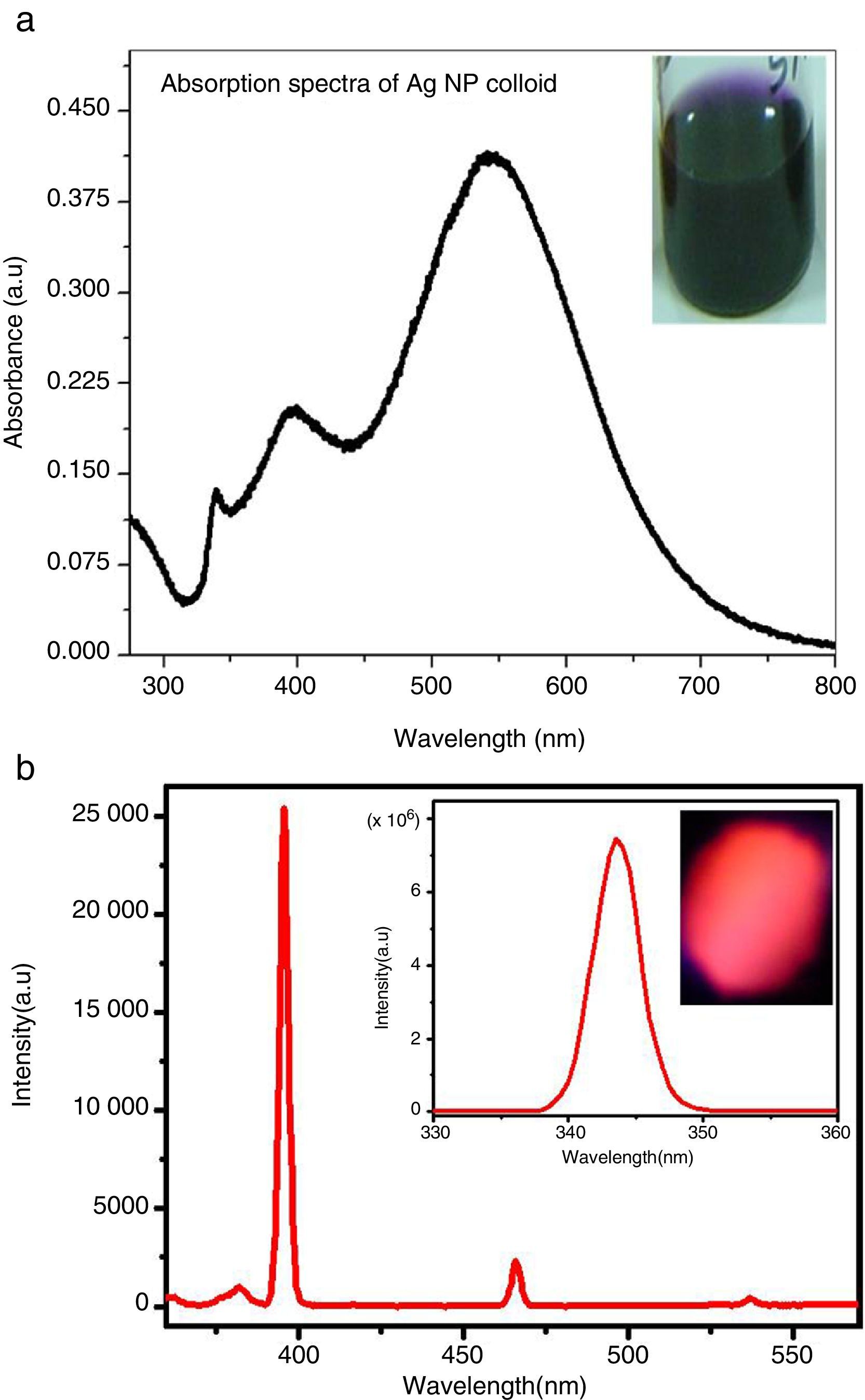
The experimental UV-visible absorption spectra of Ag NP colloid (Fig. 2a) shows a sharp out-of-plane quadrupolar peak at 338nm, an in-plane quadrupolar peak at 395nm and a broad dipolar peak centered at 542nm. The broadness of the absorption peak occurs because of the inhomogeneous damping caused by different spatial positions and random orientations of Ag NPs in the colloidal solution (Akselrod, Tischler, Young, Nocera, & Bulovic, 2010). The absorption spectra of the Ag nanoprisms cover the excitation range of GdVO4:Eu3+ as revealed by the PL excitation spectra at the red emission of GdVO4:Eu3+ (Fig. 2b), the charge transfer excitation band at 343nm and digital photograph of GdVO4:Eu3+ powder sample glowing red under UV excitation are shown in the inset of Figure 2b.
 UV-visible absorption spectrum of Ag NP colloid, the inset shows the violet colloid of Ag nanoprisms; (b) PL excitation spectra at red emission, the inset shows the charge transfer excitation band at 343nm and the digital photograph of the GdVO4:Eu3+ powder sample under UV excitation.")
(a) UV-visible absorption spectrum of Ag NP colloid, the inset shows the violet colloid of Ag nanoprisms; (b) PL excitation spectra at red emission, the inset shows the charge transfer excitation band at 343nm and the digital photograph of the GdVO4:Eu3+ powder sample under UV excitation.
Confocal mapping of the four sets of samples namely (1) Si cell+GdVO4:Eu3++AgNP; (2) Si cell+GdVO4:Eu3++PVA+Ag NP; (3) Si Cell+PVA+GdVO4:Eu3++Ag NP: (4) Si cell+PVA+GdVO4:Eu3++PVA+Ag NP was done under the excitation of a UV diode laser (375nm). The measurements on each set were done in the sequential order of layer formation, without disturbing the Si cell position on the microscope platform, keeping all the parameters the same so that the comparison of the emission intensity could be made after deposition of each layer in every set of samples and the optimum layer sequence exhibiting the maximum emission intensity could be determined.
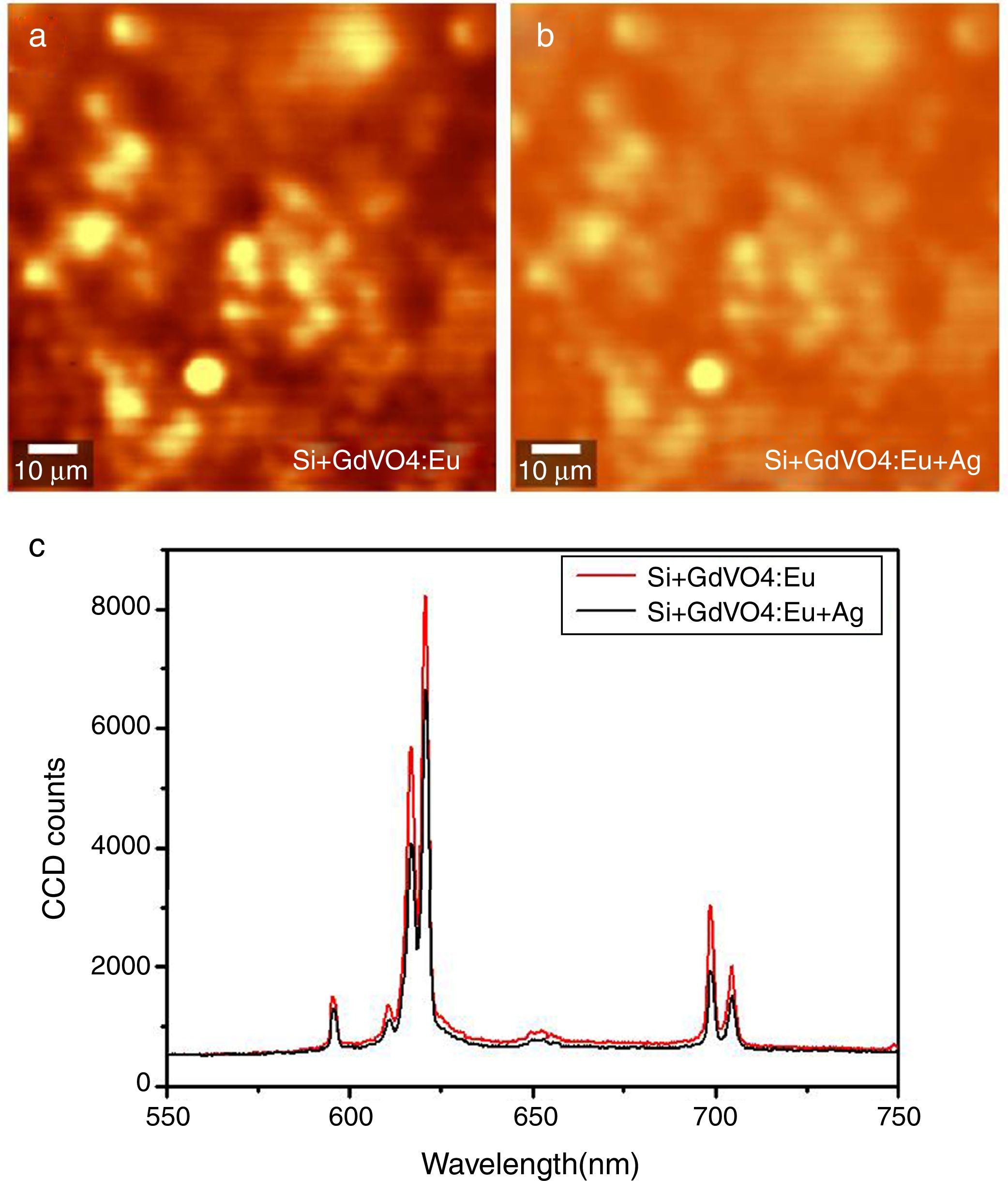
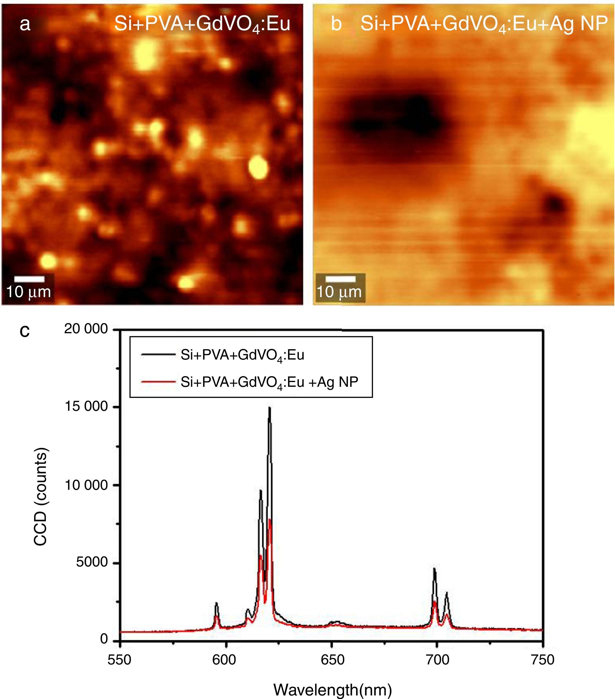
After deposition of the GdVO4:Eu3+ layer, characteristic sharp emission peaks corresponding to f-f transitions (5D0-7FJ) of Eu3+ appeared with CCD counts in few thousands. In the first set of measurement, when a comparison was made in the emission intensity of the Si cell+GdVO4:Eu3+ and Si cell+GdVO4:Eu3++Ag NPs, it was observed that on addition of Ag NPs directly on the GdVO4:Eu3+ layer, the emission was quenched to 0.82 times its original intensity as shown in the confocal fluorescence maps and the corresponding emission spectra in Figure 3.
 GdVO4:Eu; (b) GdVO4:Eu+Ag NP deposited on the Si cell; (c) corresponding spectra comparing the Eu3+ emission intensity with and without Ag NPs suggesting quenching of fluorescence.")
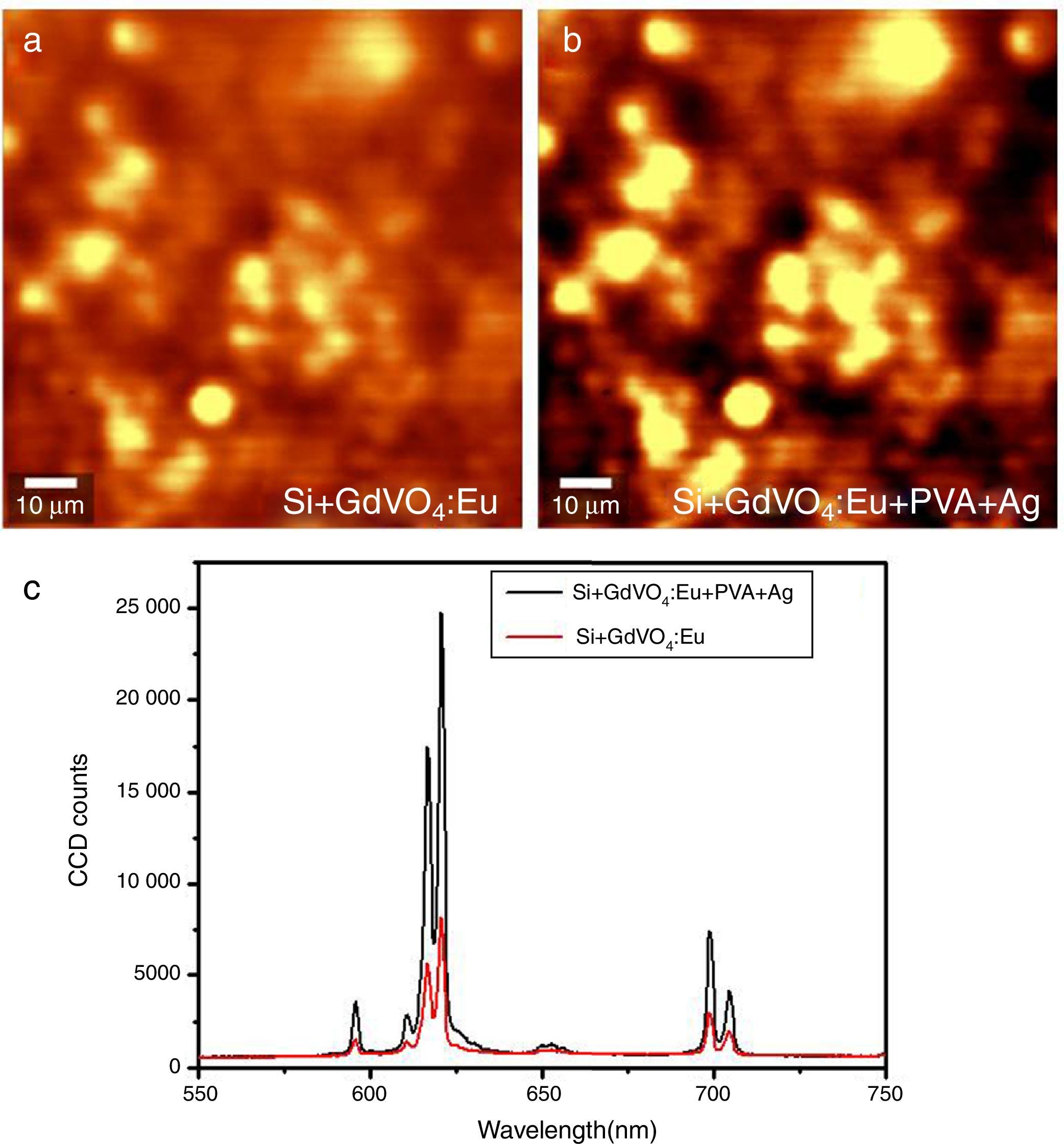
In the second set, when a dielectric spacer layer of PVA was deposited between the GdVO4:Eu3+ and Ag NPs, there occurred a significant enhancement (310%) in the emission intensity as shown in the same area fluorescence maps and spectra before and after deposition of the Ag NPs (Figure 4).
 GdVO4:Eu; (b) GdVO4:Eu+PVA+Ag NP deposited on the Si cell; (c) corresponding spectra comparing the Eu3+ emission intensity with and without Ag NPs suggesting enhancement of fluorescence.")
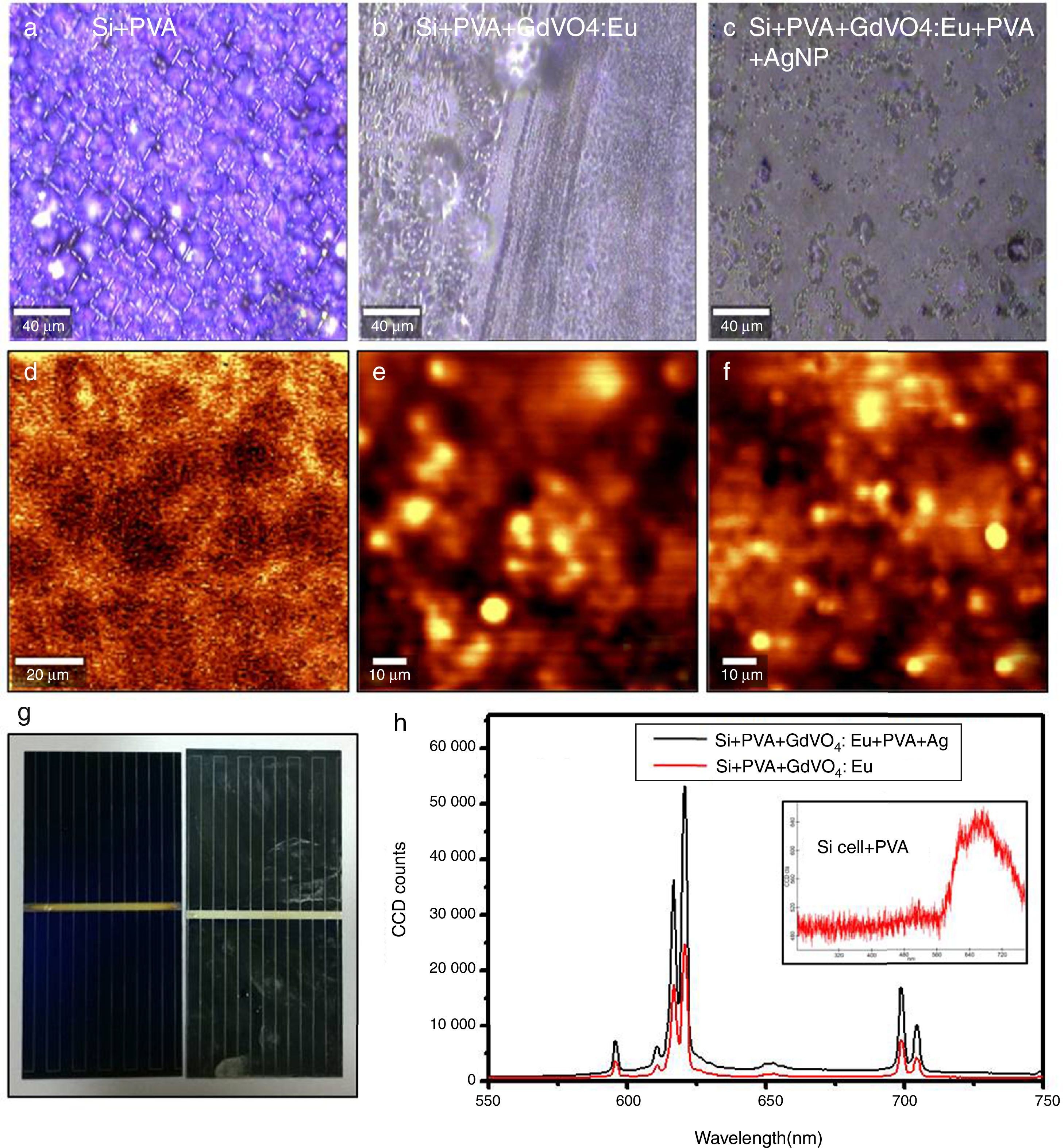
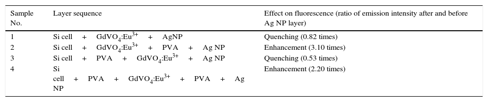
In the third set of measurements, when an additional layer of PVA was deposited between the Si cell and GdVO4:Eu3+ but no spacer layer was deposited in between the emitting species GdVO4:Eu3+ and plasmonic field generating Ag NP layer, comparison was made between the emission intensities before and after Ag NP deposition. As seen in Figure 5, it is evidently discernible that because of the addition of Ag NPs, fluorescence quenching (0.53 times its original intensity) occurs. In the same configuration, when an intervening dielectric layer of PVA was employed between GdVO4:Eu3+& Ag NPs (set 4), an excellent increment (216%) in CCD counts was observed, as shown in the confocal fluorescence maps of the same area and the emission spectra in Figure 6. Optical images and the confocal fluorescence map are shown of Si Cell+PVA (Fig. 6a and d); Si cell+PVA+GdVO4:Eu (Fig. 6b and e); Si cell+PVA+GdVO4:Eu+PVA+Ag NP (Fig. 6c and f); digital image of the bare Si cell (left) and PVA+GdVO4:Eu+PVA+Ag NP deposited on the Si cell (Fig. 6g); the corresponding spectra comparing the Eu3+ emission intensity with and without Ag NPs (Fig. 6h) suggest enhancement of fluorescence. Whereas the Bare Si cell gave emission spectra with a broad peak at 670nm with maximum 150 CCD counts (Fig. 6h, inset). The results of the PL measurements are tabulated in Table 1, which clearly elucidates the fact that an intervening dielectric layer (PVA) between the emitter layer (GdVO4:Eu3+) and Ag NP layer is essential for the enhancement of red fluorescence from GdVO4:Eu3+.
 PVA+GdVO4:Eu; (b) PVA+GdVO4:Eu+PVA+Ag NP deposited on the Si cell; (c)corresponding spectra comparing the Eu3+ emission intensity with and without Ag NPs suggesting quenching of fluorescence.")
 and (d), the Si Cell+PVA; (b) and (e), the Si cell+PVA+GdVO4:Eu; (c) and (f), the Si cell+PVA+GdVO4:Eu+PVA+Ag NP; (g), digital image of the bare Si cell (left) PVA+GdVO4:Eu+PVA+Ag NP deposited on the Si cell; (h) corresponding spectra comparing the Eu3+ emission intensity with and without Ag NPs suggesting enhancement of fluorescence, the inset shows the emission from the Si cell with the PVA layer under the same experimental conditions.")
Optical images and confocal fluorescence map of: (a) and (d), the Si Cell+PVA; (b) and (e), the Si cell+PVA+GdVO4:Eu; (c) and (f), the Si cell+PVA+GdVO4:Eu+PVA+Ag NP; (g), digital image of the bare Si cell (left) PVA+GdVO4:Eu+PVA+Ag NP deposited on the Si cell; (h) corresponding spectra comparing the Eu3+ emission intensity with and without Ag NPs suggesting enhancement of fluorescence, the inset shows the emission from the Si cell with the PVA layer under the same experimental conditions.
Effect on Eu3+ red emission of conjugation process with Ag NPs on Si cell surface.
| Sample No. | Layer sequence | Effect on fluorescence (ratio of emission intensity after and before Ag NP layer) |
|---|---|---|
| 1 | Si cell+GdVO4:Eu3++AgNP | Quenching (0.82 times) |
| 2 | Si cell+GdVO4:Eu3++PVA+Ag NP | Enhancement (3.10 times) |
| 3 | Si cell+PVA+GdVO4:Eu3++Ag NP | Quenching (0.53 times) |
| 4 | Si cell+PVA+GdVO4:Eu3++PVA+Ag NP | Enhancement (2.20 times) |
According to reports (Lakowicz, Malicka, Huang, Gryczynski, & Gryczynski, 2004), a range of preferable distance from about 40nm to about 200nm between silver nanoparticles and fluorophore lead to metal enhanced fluorescence. Particularly, for the PVA spacer layer, enhanced photoluminescence and quantum yield have been observed from J-aggregate film separated by a 100nm thick spin-coated layer of PVA (Geddes, 2013) from metal nanoparticles. For the present case, the deposited PVA spacer layer has a typical thickness of about 100nm, as measured by an optical profilometry [Stylus Profilometer (Ambios XP200)]. The investigation on four sets of samples clearly exemplify that enhancement of fluorescence occurs only when there is a PVA spacer layer between the emitter (Eu3+ in GdVO4 host) and Ag nanoprisms (sample set 2 and 4). When the PVA layer is placed in between the phosphor and Ag NPs, direct contact of the two species is averted which would have resulted in fluorescence quenching, favoring non radiative transitions from phosphor in contact with Ag NPs as observed for sample sets 1 and 3.
Metal-enhanced fluorescence occurs since highly localized electromagnetic (EM) fields generated around metal nanoparticles (MNP) by resonant excitation of conduction electrons in MNP (surface plasmons) as well as the lightening rod effect, which strictly depends on the shape of MNP, can augment fluorescence by excitation and/or emission enhancement. For such enhancement to take place, the excitation or emission spectra of phosphor must spectrally overlap with the plasmon absorption band of MNP. For the present case, the Ag NP absorption spectra show quadrupolar peaks at 338nm and 395nm and a broad dipolar peak centered at 542nm (Fig. 2a), which falls in the excitation range of the phosphor GdVO4:Eu3+ that exhibits broad charge transfer excitation band around 343nm and sharp excitation peaks at 395nm, 466nm and 537nm (Fig. 2b) because of direct excitation of Eu3+ ions. Moreover, the fluorescence enhancements and quenching observed in all the four sets of samples occur across all the characteristic 5D0–7FJ transitions in the orange to deep red-emission range of Eu3+ ions suggest an enhanced/decreased excitation of the phosphor particles from the plasmonic near field of Ag NPs due to the effect of layer sequence.
4ConclusionsIn order to achieve plasmon enhanced fluorescence on a single crystal Si solar cell, a comprehensive work on different sequence of deposition of layers of phosphor, metal nanoparticles and dielectric PVA layer on a commercial Si cell surface was carried out. The work involved chemical synthesis of shape tailored silver nanoprisms and Eu3+-doped phosphor with extended excitation range to match the absorption range of Ag nanoparticles, conjugation of Ag NP and phosphor in various configurations to ensure the best sequence for fluorescence enhancement. Confocal fluorescence mapping and spectroscopy used to investigate the same area before and after Ag NP deposition unequivocally establish plasmonic enhancement of fluorescence on the commercial Si solar cell surface. Such fluorescence enhancements (up to 310%) of Eu3+ red emission is achievable only when an intervening PVA layer separates the emitter and the metal nanoparticles whereas a PVA layer between the Si cell surface and the emitter decreases fluorescence. Direct deposition of MNPs on the emitter quenches fluorescence, with more quenching with a PVA layer between the Si cell surface and the emitter. A decrease in enhancement and an increase in fluorescence quenching because of a PVA layer on the Si cell surface may probably be caused by the absorption/scattering of the emitted photons in the interlayer. As the down shifting phosphor layer has been reported to enhance solar cell efficiency, such plasmon-enhanced red emission from vanadates on a commercial single crystal Si solar cell holds promise for further performance enhancement of silicon PV devices.
Conflict of interestThe authors have no conflicts of interest to declare.
Author S.B thanks CSIR for providing SRF fellowship and Mr. Zubair Buch for the synthesis and characterization of Ag nanoprisms.
Peer Review under the responsibility of Universidad Nacional Autónoma de México.