


The International Conference “New Trends in Immunotherapy” was held at the Hospital Universitario Puerta de Hierro (HUPH) Auditorium, in Majadahonda (Madrid) on May 20th. This event was organised by the Immunotherapy network IMMUNONET, which is funded by the SUDOE-FEDER programme (Fig. 1). This 5th edition brought together scientists and clinicians to discuss current research in the immunotherapy field.

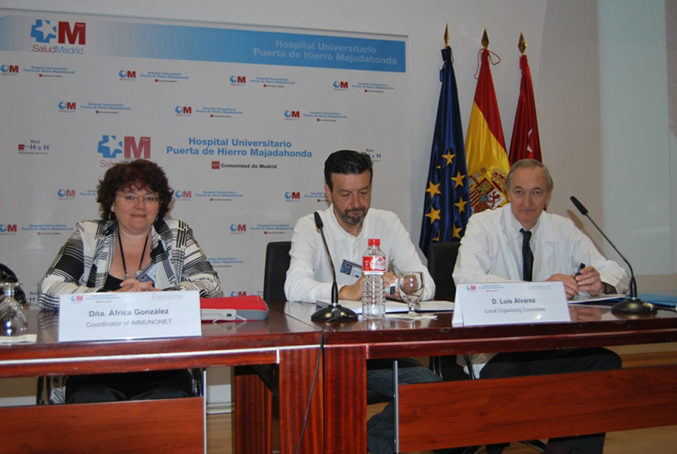
The opening event was chaired by the local organiser and core partner of IMMUNONET, Dr. Luis Álvarez-Vallina, who is Associated-Head of the HUPH Immunology Department; Dr. Fernando Díaz-Espada, Head of the HUPH Immunology Department, and Dr. África González-Fernández, Director of the Biomedical Research Centre (CINBIO) at the Universidad de Vigo, and Coordinator of the IMMUNONET network, who introduced the network (Fig. 2).
 Auditorium, in Majadahonda (Madrid) on May 20th. Right: Dr. Fernando Díaz Espada, Head of the HUPH Immunology Department. Centre: Dr. Luis Álvarez Vallina, local organiser and core partner of IMMUNONET. Left: Dr. África González-Fernández, coordinator of IMMUNONET.")
The opening event was held at the Hospital Universitario Puerta de Hierro (HUPH) Auditorium, in Majadahonda (Madrid) on May 20th. Right: Dr. Fernando Díaz Espada, Head of the HUPH Immunology Department. Centre: Dr. Luis Álvarez Vallina, local organiser and core partner of IMMUNONET. Left: Dr. África González-Fernández, coordinator of IMMUNONET.
The Immunotherapy Network IMMUNONET is a project approved under the Territorial Cooperation of the European Southwest Area Programme (SUDOE-Interreg IV B, SOE1/P1/E014). Bringing together the leading international scientists, a stable team of cutting-edge research in Immunotherapy has been established in the south west of Europe. The construction and remarkable work of the Network have been possible thanks to the great efforts of its partner members and the increasing number of collaborators and, moreover, thanks to the support of the European Regional Development Fund. The network started in 2009 with 9 core partners and 4 associated groups from France, Spain and Portugal. The groups are placed in Montpellier, Toulouse, Navarra, Porto, Lisbon, Madrid and Vigo. In just two years, the network has expanded and currently includes 24 collaborating groups and an increasing number of institutions are showing interest in participating in this initiative.
The project is structured in a virtual support centre where research groups who cover practically all the areas of Immunotherapy share resources, equipment and information with universities, public administration and private initiatives. The ultimate goal of the Network is to develop and patent immunotherapeutic agents for the treatment of infectious and autoimmune diseases, and cancer, and also initiate preclinical and clinical trials.
The training of new researchers is also one of the main priorities of the Network. These young researchers enriched their education, thanks to the easy access to some of the best experts in Immunotherapy in the world. Also, the research of these groups is providing outstanding results evident in the scientific quality of their articles and studies that the groups present at conferences worldwide.
After the presentation of the IMMUNONET network, Dr. África González introduced Dr. Fernando Díaz-Espada as the first Spanish post-doc who worked in Cambridge at the laboratory of Dr. César Milstein (Nobel Prize for Physiology and/or Medicine for the development of the hybridoma technique). Dr. Díaz-Espada paved this way the path to several other Spanish researchers who worked at Professor Milstein’s laboratory since then. Another feat ascribed to him was the introduction of the Immunotherapy in Spain where, after his return, he spent several years working in anti-idiotype vaccination for hematologic malignancies. Dr. Díaz-Espada engages with the public by remembering the history and main immunologists who contributed to the field of Immunotherapy. He pointed out that still today immunologists could be differentiated between “B cell people” and “T cell people”. He even made a demonstration by classifying the different presentations of the workshop based on the characterization he had just described. Finally, Dr. Álvarez-Vallina thanked the audience and collaborators, and the workshop started as scheduled in the programme. The presentations were focused on the role of Immunotherapy in various fields of biomedicine, such as autoimmunity, cancer, allergy, or infectious diseases.
AutoimmunitySummary: In the autoimmunity field, very interesting data were presented by Drs. Javier Hernández and Lennart Mars about several physio-pathological mechanisms that could lead to the break of the immune tolerance, including lymphopenia or crossreactivity with self-antigens. Several strategies were shown to induce the tolerance blocking the immune synapses with monoclonal antibodies directed against T cells, such as anti-CD4 (by Dr. Luis Graça) or by cell therapy using tolerogenic dendritic cells in autoimmune diseases such as multiple sclerosis and type I diabetes (by Dr. Eva Martínez Campos).
The first session started with Dr. Javier Hernández, a Spanish researcher working in the field of lymphopenia at the Institut de Génétique Moléculaire-CNRS de Montpellier (France). His presentation entitled “Factors contributing to the breakdown of peripheral T cell tolerance” showed that under lymphopenic conditions, the immune system has evolved compensatory homeostatic mechanisms that induce quiescent naive T cells to proliferate and differentiate into memory-like lymphocytes even in the apparent absence of antigenic stimulation. He hypothesized that potentially auto-reactive T cells that arrive to secondary lymphoid organs in a lymphopenic environment could differentiate and bypass the mechanisms of peripheral tolerance such as those mediated by self-antigen cross-presentation. To test this hypothesis, he used transgenic mice expressing a model antigen (HA) in the beta-cells of the pancreas as well as transgenic mice expressing an HA-specific class I and/or class II-restricted TCR. His results show that lymphopenia-driven proliferation and differentiation of potentially auto-reactive CD8+ T cells into memory-like cells are not sufficient to induce autoimmunity because they are tolerized. Induction of an organ-specific autoimmunity required the participation of antigen-specific memory-like CD4+ T helper cells. These helper cells promoted the further differentiation of memory-like CD8+ T cells into effectors in response to antigen cross-presentation resulting in their migration to the tissue of antigen expression where autoimmunity ensued. Surprisingly, this effect was not dependent on CD40–CD40L interactions but is rather mediated by IL-2. Thus, the cooperation of self-reactive memory-like CD4+ and CD8+ T cells overcomes cross-tolerance under lymphopenic conditions. This is of great importance not only to understand how autoimmunity is initiated under lymphopenic conditions but, also, to rationally design immunotherapeutic strategies against cancer.
Another member of the IMMUNONET network, Dr. Luis Graça from the Instituto de Medicina Molecular (IMM) of Lisbon (Portugal), gave a lecture on “Induction of immune tolerance with monoclonal antibodies”. Many approaches to target cancer cells are based on the use of therapeutic molecules or cells that target unique characteristics of the tumor (such as proteins only produced by the cancer cells) in order to kill them. However, the tissues of the body have evolved to protect themselves against immune-mediated damage, thus avoiding autoimmunity. As a consequence, several mechanisms are in place to reduce the effectiveness of immune-mediated killing. In addition, tumor cells tend to accumulate mutations, and as such cells that modify the proteins being targeted will have a proliferative advantage and will develop resistance to the therapeutics. An alternative approach is to reduce the action of the mechanisms that maintain immune tolerance, thus allowing more effective action of the immune system in targeting tumor cells for rejection.
Dr. Lennart Mars from the Institut National de la Santé et de la Recherche Médicale (INSERM), Toulouse (France), explained his work on “Myelin-specific T cells that co-recognise a neuronal autoantigen drive chronic inflammation in an animal model of Multiple Sclerosis”. One mechanism by which infections can break tolerance is referred to as ‘molecular mimicry’. His group has recently discovered that immunological mimicry also exists between two distinct neural antigens with the implication of self-mimicry in the CNS autoimmunity when using TCR transgenic mice expressing CD4+ T cells specific for the immunodominant epitope of MOG (myelin oligodendrocyte glycoprotein). Using these mice, the group made the paradoxical observation that their susceptibility to spontaneous EAE persisted even when the target antigen MOG was removed by genetic invalidation. A cross-reactive epitope was identified in NF-M, a member of a distinct protein family. Whereas MOG is located on the outermost lamellae of the myelin sheath, NF-M is expressed within neurons/axons. In fact, NF-M represents one of the three neurofilament subunits, which are the most abundant cytoskeletal components of large myelinated axons.
Dr. Eva Martínez Cáceres, from the Immunology Laboratory LIRAD-BST, IGTP, Germans Trias i Pujol of Badalona (Spain), gave a short overview on the possibility to treat autoimmune diseases such as Type-1 diabetes and multiple sclerosis (MS) with cell therapy. She focused on the feasibility to induce durable tolerance in relapsing-remitting MS patients by treatment with tolerogenic dendritic cells loaded with a pool of myelin peptides that are recognized by high-avidity CD4+ T cells of the patients.
Infectious diseasesSummary: Very promising data in the field of infectious diseases were presented by Luzia Teixeira and Elva Bonifácio, both collaborators of Dr. Vilanova from the Instituto de Ciências Biomédicas de Abel Salazar de la Universidad de Porto (ICBAS-UP) showing the role of regulatory T cells in a neosporosis murine model, and the implications of TLR2 and IL-10 in the development of Streptococcus agalactiae infection. Anabela Cordeiro-da-Silva, Head of the Parasite Disease Group at the IBMC - Instituto de Biologia Molecular e Celular and Facultade de Farmácia, Universidade do Porto, analyzed the role of dendritic cells in the Leishmania infection, trying to identify new targets for prevention and therapy in these pathologies.
Luzia Teixeira presented “Immunotherapy of Neospora caninum infection”, performed in collaboration with Drs. Augusto M. R. Faustino and Jocelyne Demengeot. In this work, it was shown that depletion of T regulatory cells (Treg) with an anti-CD25 monoclonal antibody, transiently increased resistance of immunodefective (IL-12/23-deficient) mice to the apicomplexan intracellular parasite Neospora caninum. This opens the possibility that transient depletion of Treg may be used to improve resistance to the infection in immunodeficient individuals.
Elva Bonifácio gave a talk about “TLR2 as a prospective target for the immunotherapy of Streptococcus agalactiae” performed with the collaborations of Pedro Madureira, Joana Alves, Adília Ribeiro, Anabela Cordeiro-da-Silva, Margarida Correia-Neves and Paula Ferreira. Severe forms of neonatal disease, as sepsis and meningitis, are caused by Streptococcus agalactiae, also known as Group B Streptococcus (GBS). The group observed that neonatal C57BL/6 wild-type (WT) mice are more susceptible to disseminated GBS infection than Toll-like Receptor-2-deficient (TLR2−/−) mice. The evaluation of the contribution of cytokines to explain these differences showed that IL-10 production through TLR2-signaling is associated with neonatal GBS susceptibility by impaired neutrophil recruitment to injured organs. These results provide evidence for the therapeutic role of IL-10 neutralization in the management of GBS infection and highlight TLR2 as a potential target molecule for immunotherapeutic strategies against GBS infections.
Finally, Anabela Cordeiro-da-Silva, gave a talk about “Distinct immunological role of Leishmania infantum on infected and bystander dendritic cells”. The group focuses its research on the interaction between DCs and the protozoan-Leishmania infantum. The distinction by flow cytometry of infected and bystander DC-populations using GFP-L infantum parasites clearly indicated the presence of two types of responding cells. While bystander DCs showed an up-regulated profile, the infected cells exhibited a more immature state. In vitro co-cultures demonstrated that infected DCs were unable to induce CD4+ and CD8+ cell activation and proliferation in opposition to bystander DCs, in a process partially dependent on the DC secretion of IL-10. The understanding of the precise role of each population may lead to the development of new forms of immunotherapy against visceral leishmaniasis.
BiotechnologySummary: The field of biotechnology was covered by Dr. Álvarez-Vallina (organizer of the event) and collaborators David Sánchez-Martín, Vanessa Alonso Camino and Laura Sanz Alcober, from the Fundación de Investigación Biomédica, Unidad de Inmunología Molecular, HUPH. They gave excellent talks about platforms of selection of antibodies directed against relevant tumor antigens, based on phage libraries or on T cells. They are also working on antibody engineering, developing new antibody molecules and a very sophisticated method of in vivo secretion of a bispecific antibody.
David Sánchez-Martín, PhD student in the group of Dr. Álvarez-Vallina gave a lecture on “In vivo selection of recombinant antibodies in tumor-bearing mice”, reporting a method for selecting recombinant antibodies (rAbs) in vivo in a tumor-bearing mouse model using a repertoire based on human VH. Monoclonal antibodies—obtained from immunized mice—are widely used in the clinics, but they face many limitations, some of which were overcome by the development of a new technology for selecting (recombinant) antibodies—phage display and the creation of immune and non-immune repertoires. There are several rAbs in different phase studies, in different formats, and against different antigens. But all the current available rAbs have been selected in vitro, against purified antigens or controlled cells populations. However, gathering evidence from the literature supports the idea of in vivo selections using peptide phage display as an alternative source of antigens in different disease models, cancer being probably the one most exploited. This in vivo approach allows the identification of antigens and epitopes that are relevant for a particular disease model that might not be accessible in other approaches (ex vivo, in vitro). Nonetheless, attempts to modify in vivo peptide phage display to the recombinant antibody field have been previously unsuccessful. They selected several antibodies that show preferential homing to the tumor. This novel rAbs may be readily adapted to any of the available strategies to improve targeting, to deliver payloads, etc. Vanessa Alonso-Camino, a PhD student in the same group talked about “Lymphocyte Display: a Novel Antibody Selection Platform based on T Cell Activation”. In this work, she described the design and testing of a mammalian cell surface display platform in T lymphocytes. The display of antibodies on the surface of T lymphocytes, as a part of a chimeric antigen receptor (CAR) mediating signaling, may ideally link the antigen–antibody interaction to a demonstrable change in T cell phenotype, due to subsequent expression of the early T cell activation marker CD69. The display of a single-chain Fv (scFv) antibody repertoire on the T cell surface in a CAR context allowed select tumor-specific antibodies in vitro.
Dr. Laura Sanz Alcober, group leader at the Unidad de Inmunología Molecular gave a lecture on “In vivo models of human angiogenesis”. Angiogenesis is a complex process implying a plethora of molecular and cellular interactions that is difficult to recapitulate in vitro. Classical in vivo models of angiogenesis relied on blood vessel formation by endothelial cells (ECs) provided by the host. In order to generate a human model of angiogenesis in vivo, with stable and functional neovessels, we have used different strategies ranging from human primary tumor xenografts to blood vessel engineering with ECs seeded in biocompatible scaffolds and inoculated into immunodeficient mice. Such vessels, engineered with ECs transduced with appropriate genes, can function as ‘cell factories’ for the in vivo production of therapeutic proteins in a variety of pathological conditions.
Finally, Dr. Luis Álvarez-Vallina presented a lecture on “Engineering human cells for in vivo secretion of antibody”. As an alternative to recombinant protein administration, ex vivo gene-modified cells may provide a novel strategy for systemic delivery of antibodies. Cell delivery vehicles may be: autologous or allogeneic, precursor or terminally differentiated cells, with targeting properties or immobilized in immunoprotective devices. Different non-hematopoietic stem cells have emerged as potential delivery vehicles, since they are easy to obtain, expand and transduce, and they exhibit prolonged life spans. They demonstrated for the first time that human mesenchymal stem cells genetically engineered to secrete a bispecific αCEA/αCD3 antibody, seeded in a synthetic extracellular matrix scaffold, and implanted in a location distant from the primary tumor, induce an effective anti-tumor response and tumor regression.
NanoimmunotherapySummary: In the last years, the development of the Nanotechnology is finding many applications in several fields (textiles, cars, energy, food, environment, medicine). Several research groups are developing several types of nanostructures, many of them with large potential in the biomedical field, either for diagnosis (in vitro or in vivo) or for human therapy. However, because they can interact more easily with the biological systems, it requires a complex and extensive previous characterization, together with biocompatibility, sterility and immunogenicity studies. There were several presentations regarding Nanotechnology, some by the group of Dr. África González, Coordinator of the IMMUNONET network, and one by the invited speaker Dr. Domingo Barber, from the Centro Nacional de Biotecnología de Madrid. These presentations touched several issues, including: the multiple possibilities of nanomaterials in the field of diagnosis, prevention and therapy, intranasal vaccines with different prototypes including Hepatitis B antigen, liberation of gamma interferon on cancer therapy, biocompatibility and immunogenicity of nanomaterials.
Tamara Lozano, master student in the group of Dr. Africa González-Fernández from the CINBIO, Universidad de Vigo, talked about “Biocompatibility of different nanoparticles on human cells”. She showed results of several metal oxide nanoparticles, analyzing aggregation, uptake, inflammation, immunogenicity and in vitro toxicity induced by different metal oxide nanoparticles (Nps) [Fe(II,III)Ox, TiOx, ZnO and CeO2] in several cell lines. The ZnO Nps were found to be highly toxic for all the cell types studied. Western blot was also used to test the ability of the different Nps to activate the complement cascade.
The immunogenicity of nanoparticles can also be used in a beneficial way in the field of vaccines. The design of effective vaccine delivery vehicles is opening up new possibilities for making immunization more equitable, safe and efficient. New ways of delivery, low doses and more stable vaccines are strongly needed and nanotechnology can help in this matter. In this work, Mercedes Peleteiro, also a master student under the supervision of Dr. África González, proposed nanoparticles as delivery structures for virus-like particle antigens using recombinant hepatitis B surface antigen (rHBsAg) as a model. In addition to in vivo test to check specific IgG antibodies using animal models, she is trying to develop in vitro models that could be used as previous screening for many prototypes of nanostructures before going to the in vivo assays. Some prototypes are showing good correlation between in vitro and in vivo results, indicating that this could be a future way to test potential nanovaccines.
Arturo Jiménez Periáñez from the Cell Biology Department, Universidad Complutense, Madrid, gave a talk about “Mesoporous silicon microparticles enhance MHC class I cross antigen presentation by human dendritic cells” showing that silicon microparticles are able to activate in vitro human dendritic cells. Finally, Domingo Barber from the Immunology and Oncology Department, Centro Nacional de Biotecnología (CNB)-CSIC, Madrid, talked about “Dimercaptosuccinic acid-coated magnetite nanoparticles for magnetically guided in vivo delivery of interferon gamma for cancer immunotherapy”. His group tested small, uniform dimercaptosuccinic acid (DMSA)-coated monodisperse magnetic nanoparticles as a delivery system for IFN-γ in mouse models of cancer. IFN-γ-loaded magnetic nanoparticles were guided to the tumor site by applying an external magnetic field, analyzing the efficiency of nanoparticle accumulation, IFN-γ release in the area of interest, and their effects on tumor development. At the tumor site it was observed nanoparticle accumulation and higher IFN-γ levels, which led to increase the infiltration of immune effector cells, an anti-angiogenic effect and tumor regression. These findings indicate that DMSA-coated nanoparticles can be used as an efficient in vivo drug delivery system.
OncologySummary: In the field of oncology, in addition to monoclonal antibodies that are finding a large number of successful applications, dendritic cells charged with tumor lysates from the same patient are being used with the purpose of increasing the immune response in cancer patients. Dr. Ignacio Melero and José Medina-Echeverz both from laboratories of the Centro de Investigación Médica Aplicada (CIMA), Navarra, showed a clinical assay using charged dendritic cells which was started in the Clínica Universitaria de Navarra with promising results. Moreover, interesting data were presented about the influence of the type of intratumoral myeloid cells in the immune response against cancer in patients going through different chemotherapy treatments. Dr. Pablo Ortiz, from the company Digna Biotech, talked about the recent track records of his company and the state of several products in clinical phases.
Other important contributions in cancer immunotherapy were given by Drs. África González y Cecilia Muñoz, from Universidad de Vigo and Instituto de Investigación Sanitaria del Hospital de la Princesa de Madrid, respectively, with promising data about the efficacy in vitro and in vivo of antibodies directed against human molecules such as HLA-DR o CCR7.
Dr. Ignacio Melero showed his work on “Biological effects of a dendritic-cell-based clinical trial”. A clinical trial performed on 24 patients with advanced cancer was presented. Patients underwent a pilot combinatorial treatment study of vaccinations with type-1 dendritic cells, GM-CSF, pegylated interferon and cyclophosphamide. Patients experienced increases in NK and T cell activity. Most patients showed high concentrations of IL-12 in post-treatment serum samples. This determined drops in circulating endothelial and tumor cells. Two patients in whom all metastatic tumors were surgically removed before treatment have not relapsed over 18 months. In light of this observation, a randomized clinical trial for colorectal cancer patients with full surgical resection of liver metastasis is ongoing at his institution and at Hospital de Navarra.
Dr. José Medina-Echeverz from the group of Dr. Pedro Berraondo presented his lecture entitled “Successful colon cancer eradication after chemoimmunotherapy is associated with profound phenotypic change of intratumoral myeloid cells”. Upsurge of regulatory T cells (Treg) in the tumor milieu has been proposed to limit the antitumor efficacy of interleukin 12 (IL-12) gene therapy. It has been showed that two drugs (cyclophosphamide [CPA] and anti-CD25 mAb), widely used to eliminate Treg in combination with IL-12, were able to eliminate intratumoral Treg and myeloid-derived suppressor cells. However, only IL-12 plus CPA achieved significant antitumor activity in mice with large established colon carcinoma. This combined treatment induced the appearance of inflammatory myeloid cells within the tumor microenvironment, an essential event to facilitate effector T cell infiltration and subsequent tumor elimination.
Dr. Pablo Ortiz presented his lecture on: “Track record Digna Biotech”, giving an interesting overview of the company. Digna Biotech is a biotechnological company dedicated to the development and exploitation of the research products generated by the Centre of Applied Medical Research (Centro de Investigación Médica Aplicada, CIMA) in Pamplona. The company works on therapeutic products, mainly to target hepatic diseases. He showed the portfolio of the products, including the state of the development of many of them. The company is centred on first clinical phases (phase I and II), leading the phase III to larger pharmaceutical companies.
Dr. África González-Fernández, presented the talk: “Fully human monoclonal antibodies directed against human class II and CD69 molecules”. In recent years, mice carrying human Ig transgenes are being generated for the production of human monoclonal antibodies as an alternative approach to the conventional use of mice or chimeric-humanized antibodies. The group used BABκ and BABκ,λ transgenic mice, which carried several genes from the human IGK (BABκ), IGK and IGL (BABκ,λ) loci, but only five human IGHV genes and the entire IGHD-IGHJ cluster linked to the human IGHM-IGHD genes. Animals were immunized with tumor lymphoid cells and several hybridomas secreting human IgM antibodies directed against human cells were obtained. One of them, obtained in collaboration with Dr. Sanchez-Madrid, recognizes the human CD69 molecule on peripheral blood-activated lymphocytes, on tumor cells of some lymphoma patients and on synovial fluid cells from arthritis rheumatoid patients, killing the inflamed cells. Another antibody, BH1 recognises HLA-class II on the surface of tumor cells from patients suffering from haematological malignancies, such as chronic and acute lymphocytic leukaemias, non-Hodgkin lymphomas and myeloid leukaemias. Interestingly, functional studies revealed that BH1 mAb recognises and kills very efficiently tumor cells from several leukaemia patients in the presence of human serum as a source of complement. Another 4 mAbs were presented, one of them able to activate bone marrow cells in vitro, expanding the granulocytic/mielocytic population.
Dr. Cecilia Muñoz from the Fundación de Investigación Biomédica, Servicio de Inmunología, Hospital Universitario de la Princesa, Madrid, gave an interesting lecture “Animal models to test the therapeutic value of the anti-CCR7 antibodies”. Her group is trying to block the dissemination of tumor lymphoid cells. They are developing monoclonal antibodies directed against CCR7, a molecule expressed in some types of leukemias and lymphomas. They use SCID mice receiving human mantle lymphoma cells to study the efficiency of mouse anti-human CCR7 antibodies in this animal model. The cells were previously transfected with a plasmid coding for luciferase, in order to analyze the evolution of the tumor in vivo by bioluminescence assays. Moreover, in some cells they will eliminate the expression of CCR7 by siRNA in order to understand the contribution of this molecule in the tumor dissemination.
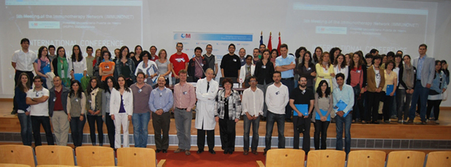
ConclusionThe quality of the presented works, the established collaborations, the mobility between participant groups and the recent incorporation of new groups to IMMUNONET indicate the great interest that this network is creating, and the huge potential as generator of knowledge and collaboration between different centres inside and outside the converge region of the Southwest of Europe (SUDOE). We would like to thank all participants for their assistance and for their interesting questions during the conference session (Fig. 3). We are already organizing the 6th meeting on Montpellier, France on 7th November 2011, hoping it would attract many scientists from the basic and applied immunotherapy fields.

We thank SUDOE-Feder (SOE1/P1/E014) for funding and support. We thank Judith Freita-Ramos for her help in the organization of this event.
The authors declare that there are no conflicts of interest in producing the manuscript.