


Prediction charts allow treatment to be targeted according to simple markers of cardiovascular risk; many algorithms do not recommend screening asymptomatic target organ damage which could change dramatically the assessment.
ObjectiveTo demonstrate that target organ damage is present in low cardiovascular risk hypertensive patients and it is more frequent and severe as global cardiovascular risk increases.
MethodsConsecutive hypertensive patients treated at a single Latin American center. Cardiovascular risk stratified according to 2013 WHO/ISH risk prediction chart America B. Left ventricular mass assessed by Devereux method, left ventricular hypertrophy considered >95g/m2 in women and >115g/m2 in men. Transmitral diastolic peak early flow velocity to average septal/lateral peak early diastolic relaxation velocity (E/e’ ratio) measured cut off value >13. Systolic function assessed by tissue Doppler average interventricular septum/lateral wall mitral annulus rate systolic excursion (s wave).
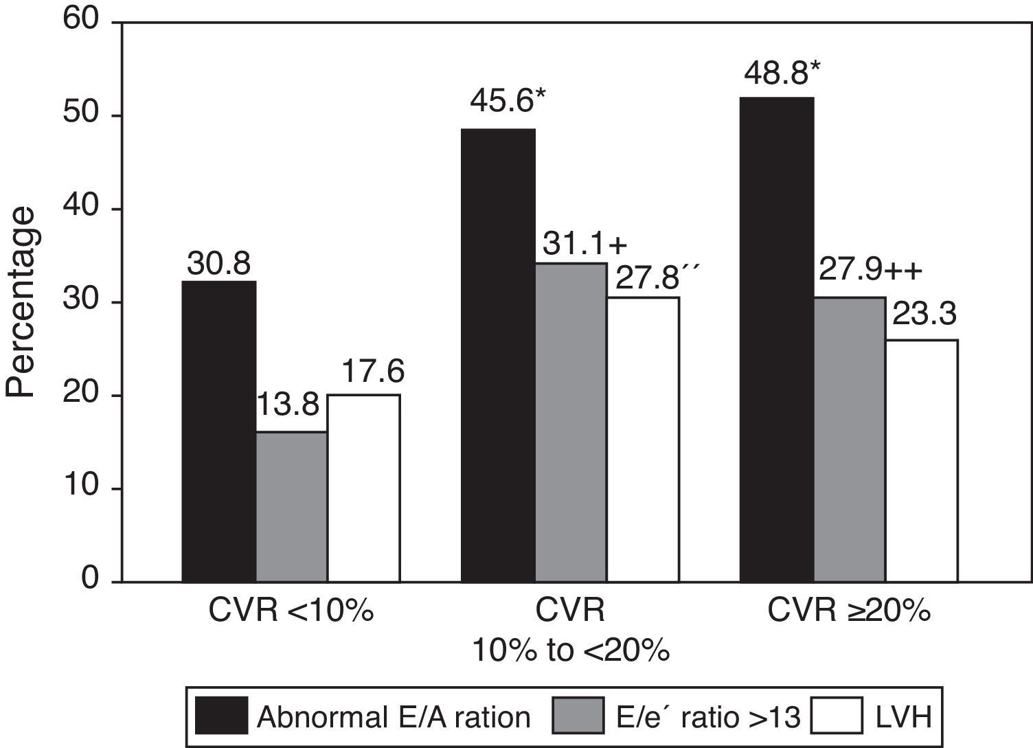
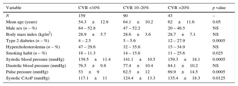
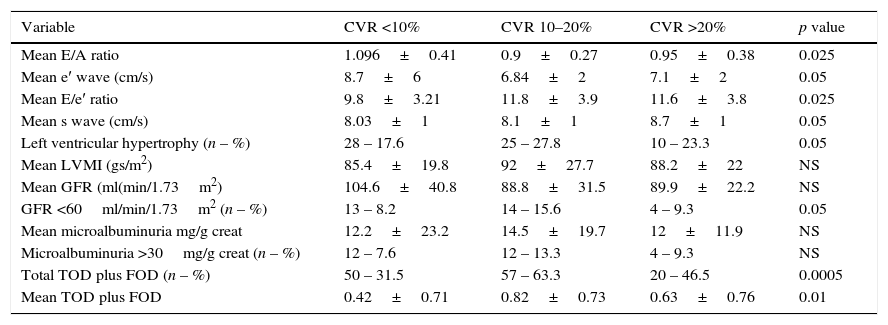
ResultsA total of 292 patients were included of whom 159 patients (54.5%) had cardiovascular risk of <10%, 90 (30.8%) had cardiovascular risk of 10–20% and 43 (14.7%) had cardiovascular risk of >20%. Left ventricular hypertrophy was detected in 17.6% low risk patients, 27.8% in medium risk and 23.3% in high risk (p<0.05), abnormal E/e′ ratio was found in 13.8%, 31.1% and 27.9%, respectively (p<0.05). Mean s wave was 8.03+8, 8.1+9 and 8.7+1cm/s for low, intermediate and high risk patients, respectively (p<0.025).
ConclusionsTarget organ damage is more frequent and severe in high risk; one over four subjects was misclassified due to the presence of asymptomatic target organ damage.
Las tablas de riesgo cardiovascular dirigen el tratamiento según marcadores clínicos sencillos. Muchos algoritmos no recomiendan el cribado rutinario del daño de órgano blanco asintomático que podría cambiar drásticamente la estratificación.
ObjetivosDemostrar que el daño en órgano blanco es altamente prevalente en el bajo riesgo cardiovascular y más frecuente y severo en la medida en que este aumenta.
Material y métodosUn total de 292 pacientes hipertensos consecutivos no tratados en un único centro latinoamericano. Riesgo cardiovascular estratificado según Guía 2013 OMS/ISH América B. Masa ventricular izquierda evaluada por método de Devereux, hipertrofia ventricular izquierda >95g/m2 mujeres y >115g/m2 hombres. Se midió relación velocidad pico diastólico transmitral con doppler y velocidad diastólica precoz septal y lateral del anillo mitral con doppler tisular (relación E/e′), valor de corte >13. Función sistólica evaluada por doppler tisular como tasa de excursión tabique interventricular y pared lateral (onda s).
ResultadosUn total de 159 pacientes (54,5%) presentaron riesgo cardiovascular <10%; 90 (30,8%) riesgo cardiovascular entre el 10% y el <20% y 43 (14,7%) presentaron un riesgo cardiovascular >20%. La hipertrofia ventricular izquierda en 17,6% pacientes fue de bajo riesgo, en el 27,8% de riesgo intermedio y en el 23,3% de alto riesgo (p<0,05), con relación E/e′ anormal 13,8; 31,1 y 27,9%, respectivamente (p<0,05). La onda s promedio fue de 8,03+8; 8,1+9; y 8,7+1cm/seg para riesgo bajo, intermedio y alto, respectivamente (p<0,025).
ConclusionesEl daño en órgano blanco fue más frecuente y severo en alto riesgo; uno de cada 4 sujetos fue clasificado erróneamente debido a presencia de daño en órgano blanco subclínico.
