


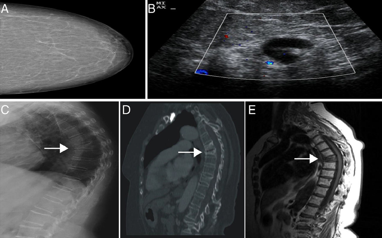
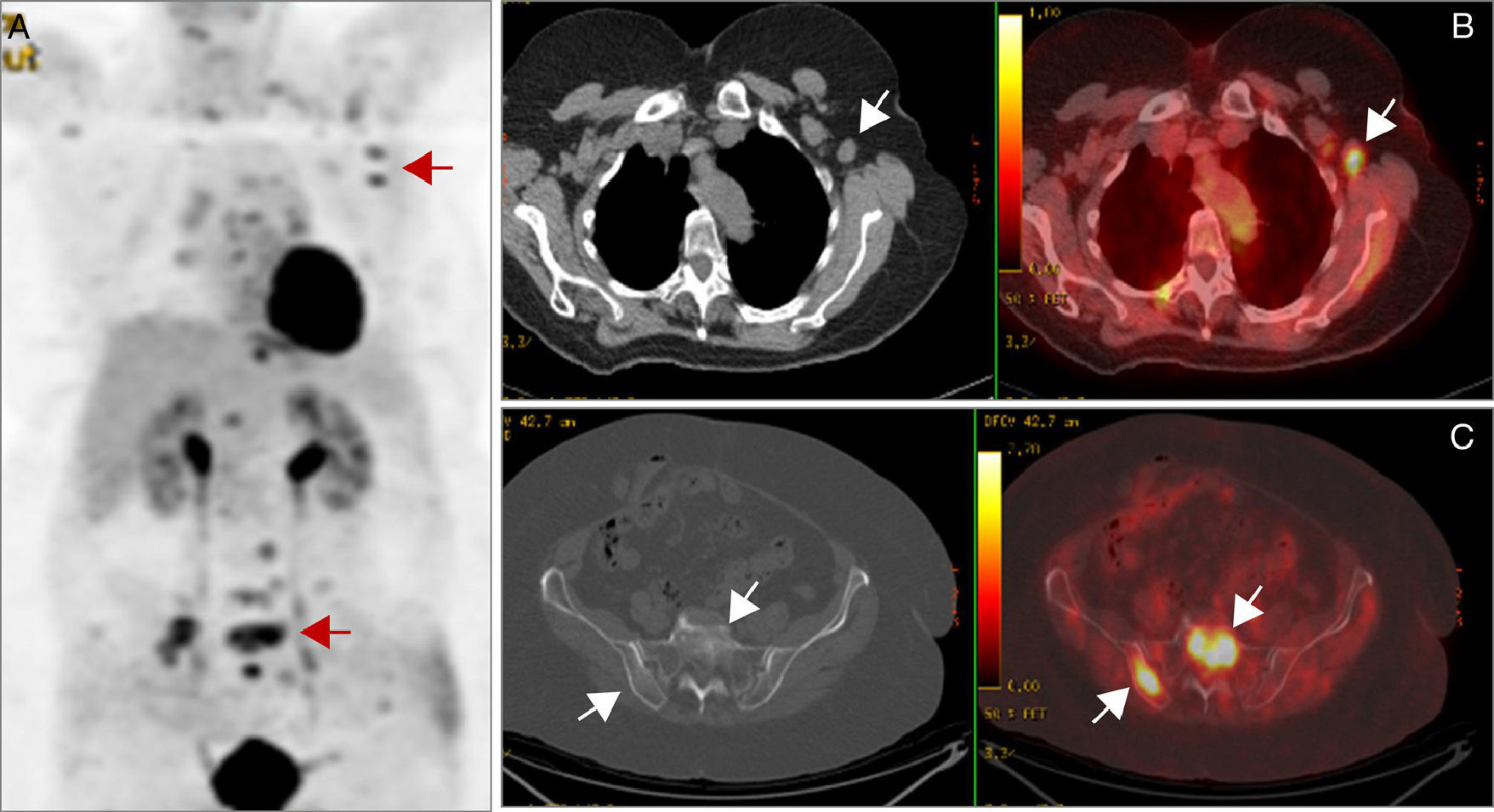
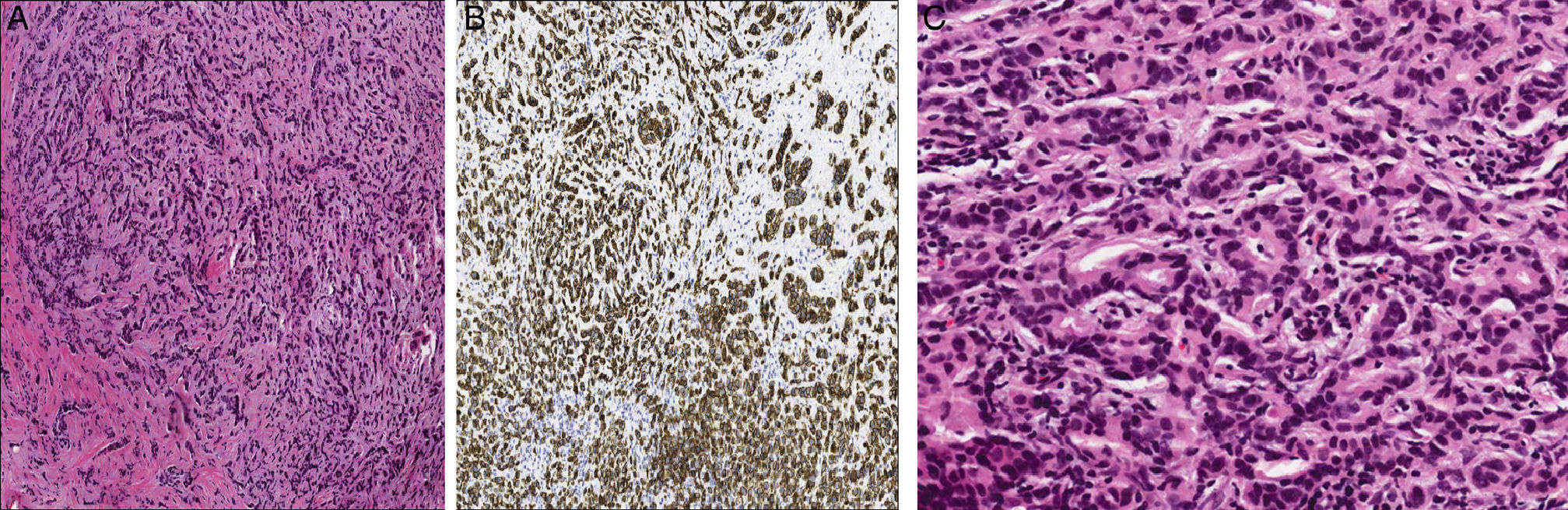
A 68-year-old female patient with spondylolysis, fibromyalgia, osteoporosis and recent BI-RADS I (negative mammogram malignancy without nodules or calcifications) mammography screening presented lumbar pain after physical overexertion, with a vertebral crush fracture on D7 and normal laboratory tests (tumor, bone metabolism markers and acute phase reactants). Subsequently, given the progressive worsening, an extension study with bone scintigraphy (BS), computed tomography (CT) and magnetic resonance imaging (MRI) was performed, showing lesions suggestive of an infiltrative process in the D6, D7, D10 and L5 vertebral bodies and nonspecific left axilla lymphadenopathy, without a clear primary origin (Fig. 1) Finally, she was studied using positron emission tomography (PET) to identify a small nodule in the lateral and deep region of the left breast, with pathology findings suggesting a metabolic origin (Fig. 2), which led to an ultrasound-guided biopsy and a histological diagnosis of moderately differentiated infiltrating ductal carcinoma (Fig. 3). Hormonal treatment and lumpectomy of the nodule as well as left axillary lymphadenectomy was performed with a good outcome.
 Mammography BIRADS I. (B) Adenopathy with cortical thickening suspicious of metastasis. (C–E) A D7 vertebral fracture with a sclerotic pattern is seen in the sagittal CT and a hypointense signal on MR imaging T1.")
 Pathological uptake in the GO level of vertebral bodies and at the level of left axillary lymphadenopathy. (B and C) A positive correlation between bone and lymph nodes with an increase of metabolic activity in the PET is observed.")
 Hematoxylin–eosin (4×); tumor cells with hyperchromatic nuclei are observed. (B) Immunohistochemical staining (4×) cells with expression of E-cadherin, surrounded by a desmoplastic stroma. (C) At higher magnification (20×), other areas of the cylinder forming gland precursors infiltrating the interstitium, which is fibrous and shows an inflammatory reaction, may be seen.")
(A) Hematoxylin–eosin (4×); tumor cells with hyperchromatic nuclei are observed. (B) Immunohistochemical staining (4×) cells with expression of E-cadherin, surrounded by a desmoplastic stroma. (C) At higher magnification (20×), other areas of the cylinder forming gland precursors infiltrating the interstitium, which is fibrous and shows an inflammatory reaction, may be seen.
The spine is the most common site of bone metastases; it is estimated that about 10% of cancer patients develop spinal metastases (>50%multilevel metastases), especially associated to carcinoma of the breast, prostate and lung. While up to 36% of spinal lesions may be asymptomatic, low back pain secondary to a pathologic fracture is the predominant symptom.1,2
Occult breast cancer consists in the appearance of a metastatic axillary lymph node, without a clinically and radiologically primary breast tumor; this is rare, and described in 0.3%–0.8% of all breast tumors.3,4
The first images, during the initial approach, are based on plain x rays. However, in most cases these findings are often nonspecific, making the use of other complementary imaging tests such as GO, CT, MRI and PET for providing great help in determining the cause of vertebral involvement.5Of these, PET will identify lesions with an abnormal metabolism without an objective anatomic abnormality (sensitivity 62%–100% and specificity 96%–100%).6 As for hidden breast cancer, the detection of such small lesions allows for the diagnosis when other conventional imaging tests do not lead to identification.7,8
Ethical ResponsibilitiesProtection of people and animalsThe authors declare that this study did not perform experiments on humans or animals.
Data confidentialityThe authors declare that they have followed the protocols of their workplace on the publication of data from patients, and all patients included in the study have received sufficient information and gave written informed consent to participate in the study.
Right to privacy and informed consentThe authors state that patient data does not appear in this article.
DisclosuresThe authors have no disclosures to make.
Please cite this article as: Ramírez Huaranga MA, Salas Manzanedo V, Huertas MP, Torres Sousa Y, Ramos Rodríguez CC. Dolor lumbar como única manifestación de un cáncer oculto de mama, utilidad de la tomografía por emisión de positrones. Reumatol Clin. 2015;11:118–120.


