


Hemangiomas are the most frequent benign solid tumors in the liver and spleen.1–5 They are usually found as single tumors in one organ. Multiple hemangiomas in the spleen and liver are an exceptional finding. We present a new case and discuss the therapeutic options.
A 50-year-old woman complained of mild abdominal pain in the epigastrium. Abdominal examination was normal. Lab work included: full blood panel, hepatic profile and tumor markers (CEA and CA19-9), which were normal. On abdominal ultrasound, 3 round images were observed that were well-defined and hyperechoic, 2 of which were in segment viii measuring 41 and 37mm, respectively, and another in segment vii measuring 11mm. These images were consistent with hemangiomas. Several lesions were observed in the splenic parenchyma of up to 2.5cm with similar characteristics and equal pathological significance to those in the liver parenchyma.
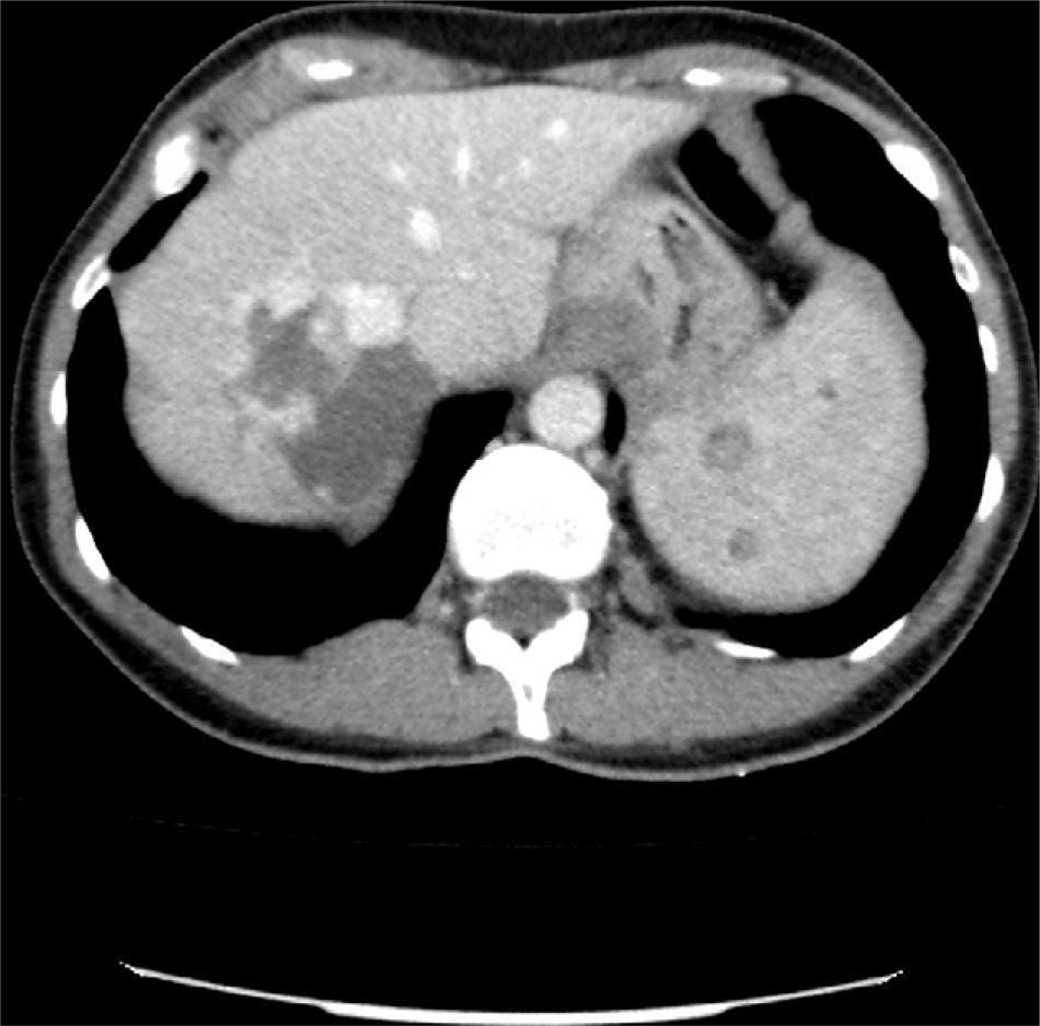
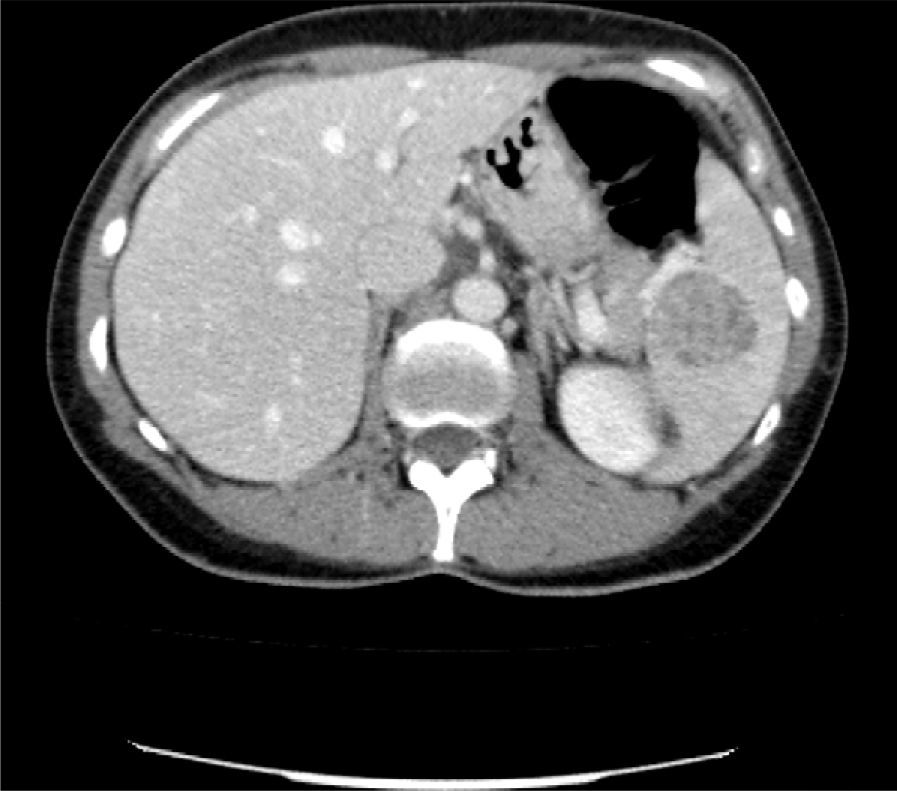
On CT, the liver showed normal size and morphology. There was evidence of several lesions dispersed throughout both liver lobes. The larger ones were located in segment viii and measured 50 and 40mm, respectively. These lesions were hypodense and presented protruding peripheral uptake of the contrast material, compatible with hemangiomas (Fig. 1). Other hepatic lesions were identified with rounded and hypodense morphology that presented diameters smaller than 1cm, which were located in segments ii (2), iv (1), vi (3) and vii (1). This latter seemed to correspond with the lesion seen on ultrasound as hyperechoic. One of the lesions in segment vi presented peripheral uptake of the contrast material. All the lesions corresponded with hemangiomas (Fig. 2).


The spleen measured 10cm. Seven lesions were observed in the splenic parenchyma, with rounded morphology and well-outlined profiles. These lesions were of different sizes: the largest had a maximum diameter of 3.5cm and the smallest 0.5cm. The lesions were hypodense and presented with heterogeneous uptake of the contrast material. These findings, together with the previously mentioned ultrasounds, were compatible with hemangiomas.
With the diagnosis of multiple asymptomatic liver and spleen hemangiomas, the patient was scheduled for regular follow-up.
Hemangiomas are the most frequent benign solid tumor of the liver.1,2,4 They are congenital vascular lesions that occur because of a hamartomatous proliferation of endothelial vascular cells that are not potentially malignant.1 There are 2 subtypes: cavernous (80%), which can grow and cause symptoms; or capillary, which are smaller hemangiomas that are more frequently multiple and usually asymptomatic.1 The diagnosis is usually incidental.4 CT and MRI are the most effective diagnostic methods.4,6,7 Hepatic hemangiomas should only be resected if there is doubt of the diagnosis, symptoms or complications.1,4 In complicated or very symptomatic patients with multiple lesions, surgical resection can be extremely complex. Another therapeutic option that is sometimes used in these patients is percutaneous embolization of the hepatic artery.2,4
Hemangiomas are the most frequent benign splenic primary tumor.3,5–7 Their prevalence in autopsies ranges between 0.03 and 14%.3,5 Hodge described the first surgical removal in 1895. Since then, only 100 cases have been reported.5 They are usually single, small, asymptomatic lesions and are 9 times more frequent in women.3 Diagnosis is usually incidental and the key to treatment is reaching a correct diagnosis in order to make proper therapeutic decisions.5 The ideal treatment for splenic hemangioma has not been clarified.5 Certain authors advocate splenectomy due to the possible risk of malignization toward angiosarcoma, affirming that this occurs more frequently in large hemangiomas or when there is diffuse splenic involvement. But the actual possibility of malignization seems remote and is not well defined.5 Other authors defend splenectomy due to possible spontaneous rupture especially in lesions that are large or symptomatic.5,8 The series by Willcox et al., which is the largest published series with 32 splenic hemangiomas, recommends monitoring small asymptomatic lesions, although they do not comment on cases with multiple hemangiomas.5 The surgical options included partial or total splenectomy.3 Percutaneous embolization is a therapeutic option that is also used.3 Several drug treatments used to produce the regression of hepatic or splenic hemangiomas have not been shown to be effective.3
The presence of multiple hemangiomas in the liver and spleen in adults is very rare. They may appear as diffuse hemangiomatosis in the so-called angiomatous syndromes.6,7 We have only found two cases similar to the case that we present in the literature after a database search (Pubmed) using the keywords hemangioma, spleen and liver.2,3 The treatments used in these two cases were: percutaneous embolization of the liver and spleen hemangiomas and later splenectomy in one case, and splenectomy and observation of the hepatic hemangiomas in the other patient.2,3 Since our patient was asymptomatic and the diagnosis was certain, we decided to follow the criteria put forth by Willcox in splenic hemangiomas and accepted in liver hemangiomas, which involves periodical observation of these hemangiomas.5
Please cite this article as: Ramia Ángel JM, Gijón de la Santa L, de la Plaza Llamas R, Quiñones Sampedro J, García-Parreño Jofre J. Hemangiomas múltiples hepáticos y esplénicos. Cir Esp. 2014;92:211–212.
