


Tracheal bronchus is considered a rare, congenital anomaly, which implies the abnormal origin of a bronchus. When related to repetitive infections the bronchus must be resected, usually via an open procedure.
ObjectiveThe aim of this paper is to present the case of a patient with tracheal bronchus of the upper right lobe who presented with repetitive pneumonias. Additionally, this text intends to expose the methodology for its diagnosis and surgical resolution through a thoracoscopic lobectomy.
Clinical caseOne year old female patient who presented with the disorder at two months of age. The patient presented with constant coughing and persistent fever alongside repetitive pneumonias in the upper right lobe. In order to discard the possibility of gastroesophageal reflux, a bronchoscopy and a panendoscopy of the digestive tube were conducted. The aforesaid procedure demonstrated the existence of a tracheal bronchus located in the right lobe, with functional bronchial segmentation. With these findings and due to the presence of repetitive infections, an apical right lobectomy was performed through a thoracoscopy, with favourable results.
ConclusionsTracheal bronchus is a rare anomaly that on many occasions is asymptomatic; nonetheless, when related to repetitive infections, a lobectomy must be carried out to avoid further pulmonary damage. This can be done through a thoracoscopy, as was the case with our patient. When treating these patients, it is worth considering they tend to have a different anatomy and to consider the ease at which they can sustain severe inflammation due to repetitive infections.
El bronquio traqueal es una anomalía congénita poco frecuente que implica el origen anormal de un bronquio. Cuando se asocia con infecciones de repetición se debe resecar este, lo cual habitualmente se hace mediante procedimientos quirúrgicos abiertos.
ObjetivoEl objetivo de este trabajo es presentar el caso de un paciente pediátrico con bronquio traqueal del lóbulo superior derecho, con neumonías de repetición, que fue tratado mediante una lobectomía toracoscópica.
Caso clínicoPaciente femenina de un año de edad, que inició su afección a los 2 meses de vida con tos productiva en accesos y persistente, con neumonías de repetición en el lóbulo superior derecho. Para descartar reflujo gastroesofágico, se practicó broncoscopia y panendoscopia de tubo digestivo, en donde se documentó la emergencia del bronquio para el lóbulo superior derecho por arriba de la carina; se practicó una tomografía axial computada en donde se encontró la emergencia del bronquio apical derecho 2cm por arriba de la carina principal, con segmentación bronquial funcional. Con estos hallazgos, y al estar relacionado con infecciones de repetición, se llevó a cabo una lobectomía apical derecha por toracoscopia sin complicaciones.
ConclusionesEl bronquio traqueal es una anomalía rara, que en muchas ocasiones es asintomática pero cuando se asocia a infecciones de repetición debe realizarse una lobectomía para evitar infecciones pulmonares. Es posible el abordaje toracoscópico pero se debe tener en cuenta que estos pacientes presentan una anatomía diferente, con mayor inflamación debido a los procesos infecciosos de repetición.
Tracheal bronchus refers to any portion of the airway arising from the trachea above the main carina.1 Its incidence ranges from 0.1% to 2% of the population.2,3 In most cases it is found incidentally during an endoscopic or radiological study.4,5 Most patients do not have symptoms and therefore do not require treatment; however, in cases of recurrent pneumonia of the anomalous lobe, the treatment of choice is to resect it.2,6 To date, surgical treatment has been undertaken in the traditional fashion through open thoracic approaches. There are no reports of thoracoscopic lobe resection in patients with tracheal bronchus.7
The aim of this paper is to present the case of a girl with a right tracheal bronchus and recurrent pneumonia, the diagnostic methodology and successful treatment by apical right lobectomy through thoracoscopy.
Clinical caseThe patient was a one-year old female, product of a multiple pregnancy, delivered by caesarean at 32 weeks’ gestation. The patient's birth weight was 1650g; she was kept in the neonatal intensive care unit, did not require assisted ventilation, and was discharged aged one month.
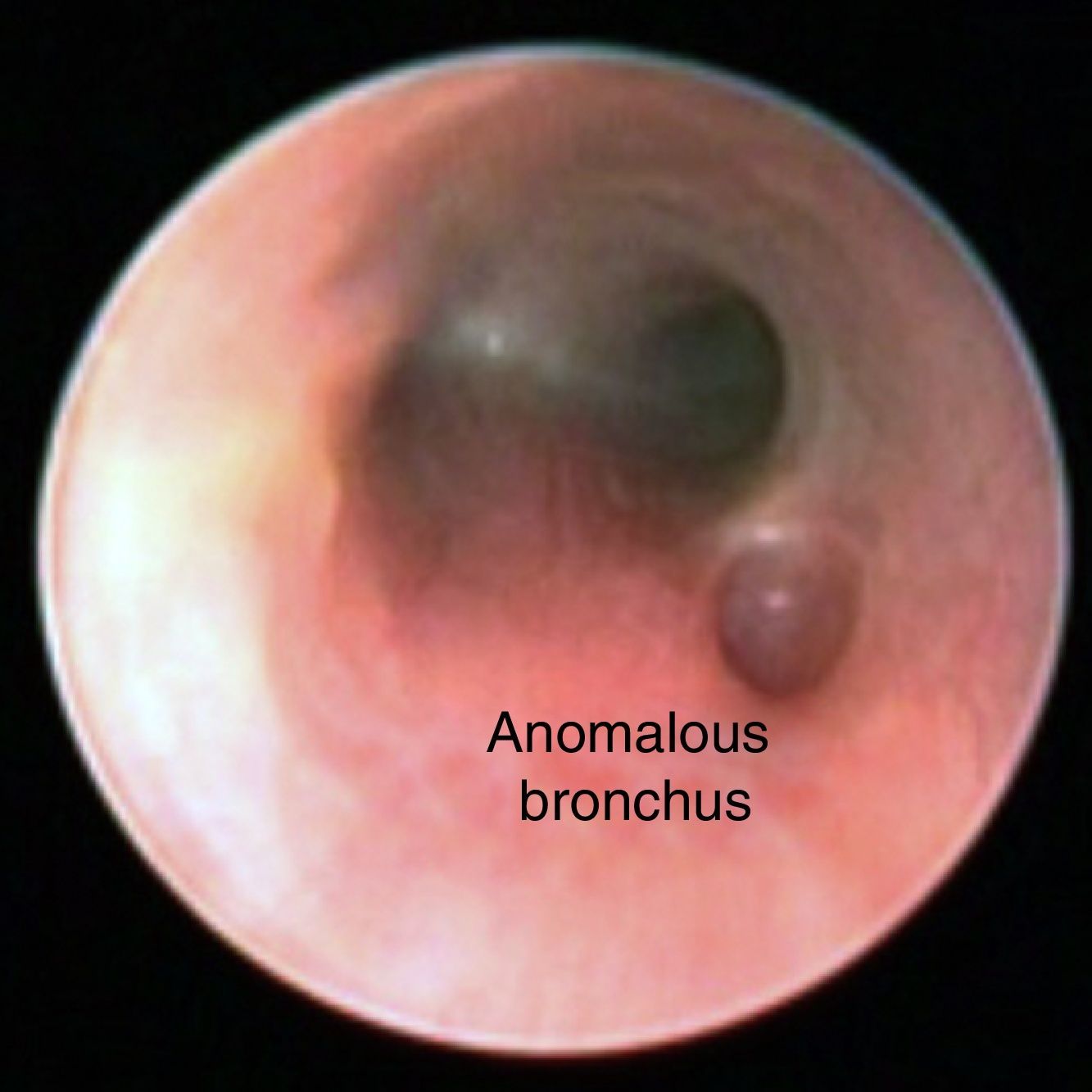
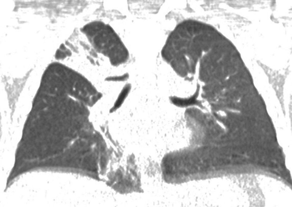
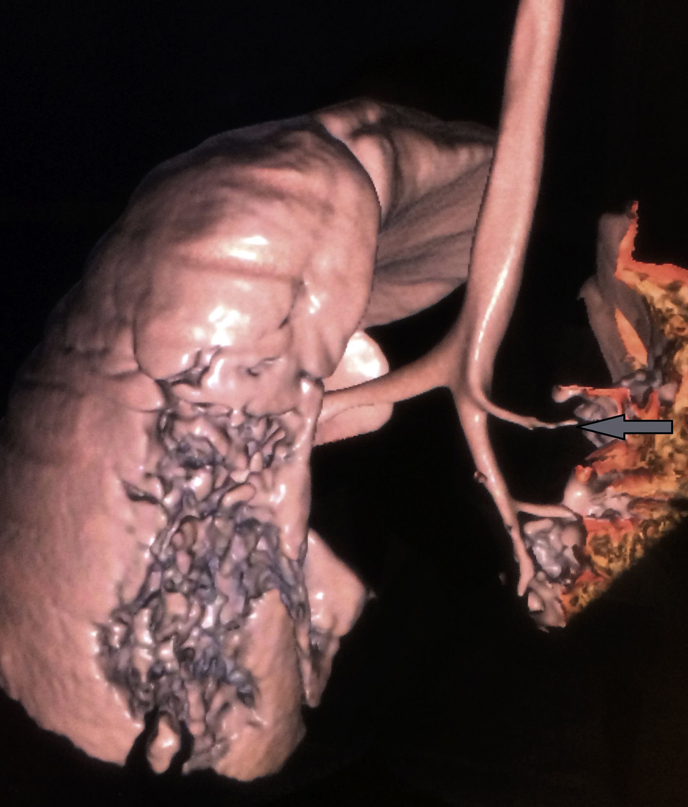
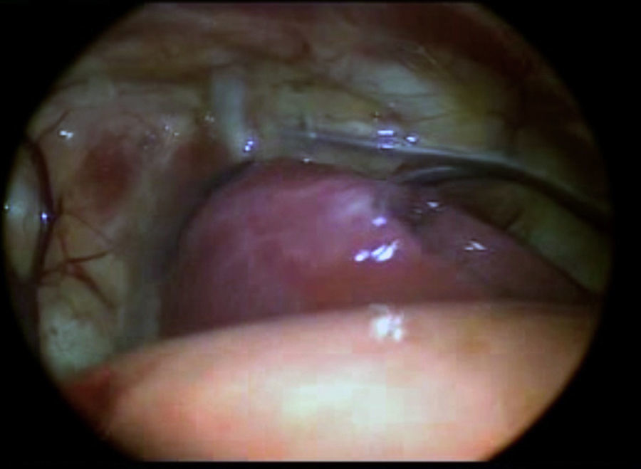
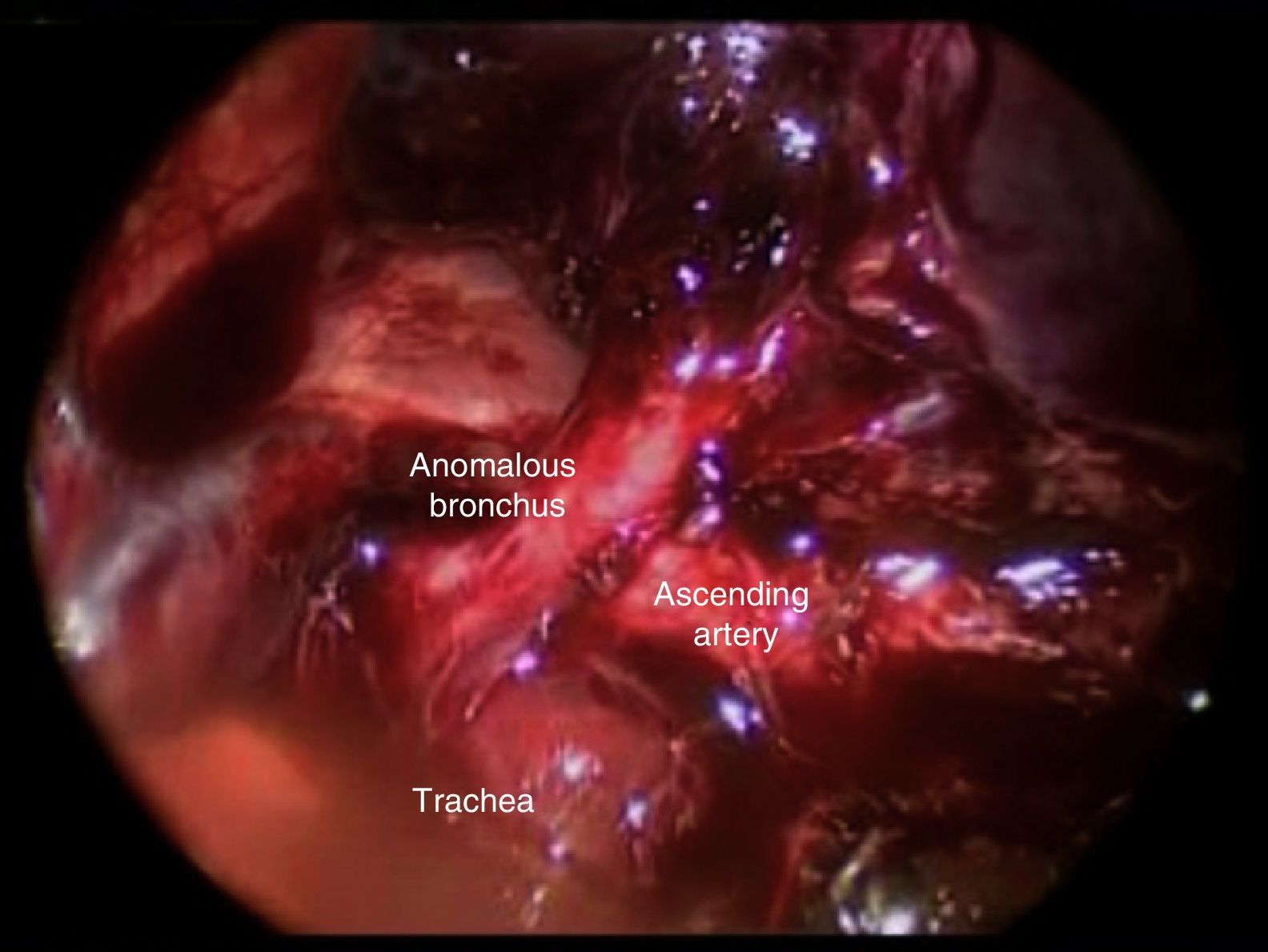
Her current condition started at the age of 2 months, with spells of productive coughing. She was treated with different antibiotic regimens, and the presence of repeated right apical pneumonias was documented; for this reason, because we considered that this might be associated with gastro-oesophageal reflux, she underwent bronchoscopy and digestive panendoscopy. Endoscopic study of the bronchi showed the bronchus arising in the right upper lobe of the lateral face of the trachea, 1cm above the main carina (Fig. 1). In order to determine the lobe pattern that corresponded with this bronchus, we performed axial computed tomography which revealed correspondence of this bronchus with the right upper lobe (Figs. 2 and 3). After a tracheal bronchus with functional bronchial segment was diagnosed, the patient was scheduled for apical right lobectomy through thoracoscopy. Bronchoscopy and aspirate were performed preoperatively, and abundant purulent material was found, which was sent for culture and reported 100,000 CFU Haemophilus influenza. The patient was then placed in the left lateral decubitus position and a 4 port approach was used. The first port, of 5mm, was for the optic, 2cm below the tip of the scapula, using a 5mm, 30° lens; 2 ports in the 2nd and 6th intercostal space, posterior axillary line of 5mm and one in the 2nd intercostal space, anterior axillary line of 5mm. CO2 was insufflated at a pressure of 5mm and flow of 0.5l/min to collapse the lung. At the start of the exploration, a consolidated upper lobe was found (Fig. 4); the ascending pulmonary artery was dissected and the bronchus of the right upper lobe (Fig. 5), the fissure was released using a harmonic scalpel, the ascending pulmonary artery was cut and the bronchus stapled with a lateral 30mm endoscopic stapler. The lobe was retracted towards the anterior part of the thorax and the two remaining pulmonary arteries were identified then sealed and cut. Finally, the 3 branches of the pulmonary vein for this lobe were dissected in the same manner. The patient made adequate progress, postoperative radiography showed appropriate lung expansion. The chest tube was removed on the fourth day and the patient was discharged on completion of the antibiotic treatment. The patient had remission of her infectious symptoms over a 12-month follow-up. The histopathological report showed inflammatory changes secondary to infectious processes.


Tracheal bronchus can present as an anatomical variant in up to 2% of the population.8 There are 2 theories as to how it forms; it is a tracheal bud that does not regress, or it is due to the bronchial mesenchyma implanting inside the trachea.8
There are 3 types of tracheal bronchus; the first is only a tracheal diverticulum, the second is a tracheal bronchus with an apical segment, and the third has a functional bronchial segment.3
Our patient had the latter. Likewise, this girl's anomaly fulfils all the requirements to be considered a true tracheal bronchus or pig bronchus, since the upper right lobe arose from the trachea and the main right bronchus provided branches for the middle and upper lobe; this type of anomaly only occurs in 0.2% of the general population.4–9 The anomaly can present in isolation, as with our patient, or can be associated with other disorders, such as pectus excavatum, cystic adenomatoid malformation and Fallot's tetralogy, the latter is the heart disease most often associated with tracheal bronchus, and therefore it is important to rule it out.4–10
A tracheal bronchus can be asymptomatic or can manifest as recurrent apical pneumonia, as in our patient.
Diagnosis can be made by bronchoscopy,11 as in the case reported here, where it was used to investigate persistent right apical pneumonias and found the accessory bronchus arising from the right lateral wall at less than 2cm from the main carina of the trachea.11
There are 2 types of tracheal bronchus; one is termed supernumerary and the other displaced.3–9 The latter was found in our case.
All can be confirmed by computed axial tomography – an excellent aid in assessing the airway and the emergence of the bronchi.8
Tracheal bronchus will be treated according to the severity of symptoms; most cases can be treated conservatively, but patients with recurrent pneumonia require surgical resection of the affected pulmonary lobe.10
This treatment has been performed by open thoracotomy. The first pulmonary lobectomies using minimally-invasive approaches were performed by Rothenberg in 1995.12 Since then, several papers have been published that demonstrate the feasibility of performing pulmonary lobectomies in children by thoracoscopy, principally for treating cystic adenomatoid disease, congenital lobar emphysema and severe bronchiectasis, which have reduced the morbidity associated with the large wounds produced in open thoracotomies, and result in better recovery with less pain.
However, none of these publications mention the procedure in paediatric tracheal bronchus patients, as we report in this paper.12–14
There are only 2 reports of thoracoscopic right apical lobectomies in adult tracheal bronchus patients associated with lung cancer.15,16
This paper reports a right apical lobectomy in a paediatric patient with displaced tracheal bronchus performed through thoracoscopy with an adequate postoperative outcome. However, the following aspects should be considered when performing this operation on these types of patients:
- 1.
A history of recurrent pneumonia favours a greater number of pleuropulmonary adherences, which will make the operation more difficult and increase the risk of haemorrhage.
- 2.
These patients can have associated vascular malformations.15
None of these conditions prevented us from performing the minimally-invasive procedure in the case we present in this paper.
ConclusionThis report demonstrates the possibility of undertaking minimally invasive pulmonary lobectomies in paediatric tracheal bronchus patients, achieving good outcomes and an efficacious cure of the condition.
Ethical disclosuresProtection of human and animal subjectsThe authors declare that no experiments were performed on humans or animals for this study.
Confidentiality of dataThe authors declare that they have followed the protocols of their work centre on the publication of patient data.
Right to privacy and informed consentThe authors declare that no patient data appear in this article.
Conflict of interestThe authors have no conflict of interests to declare.
Please cite this article as: García-Hernández C, Carvajal Figueroa L, Celorio Alcántara Á, Landa-Juárez S, Salinas Hernández E. Lobectomía toracoscópica para el tratamiento del bronquio traqueal. Reporte de un caso pediátrico. Cir Cir. 2017;85:557–561.