
We present the development of a tool for the automatic detection of microaneurysms and its clinical evaluation. The intention of this tool is to facilitate the diagnosis of diabetic retinopathy in general screening programs.
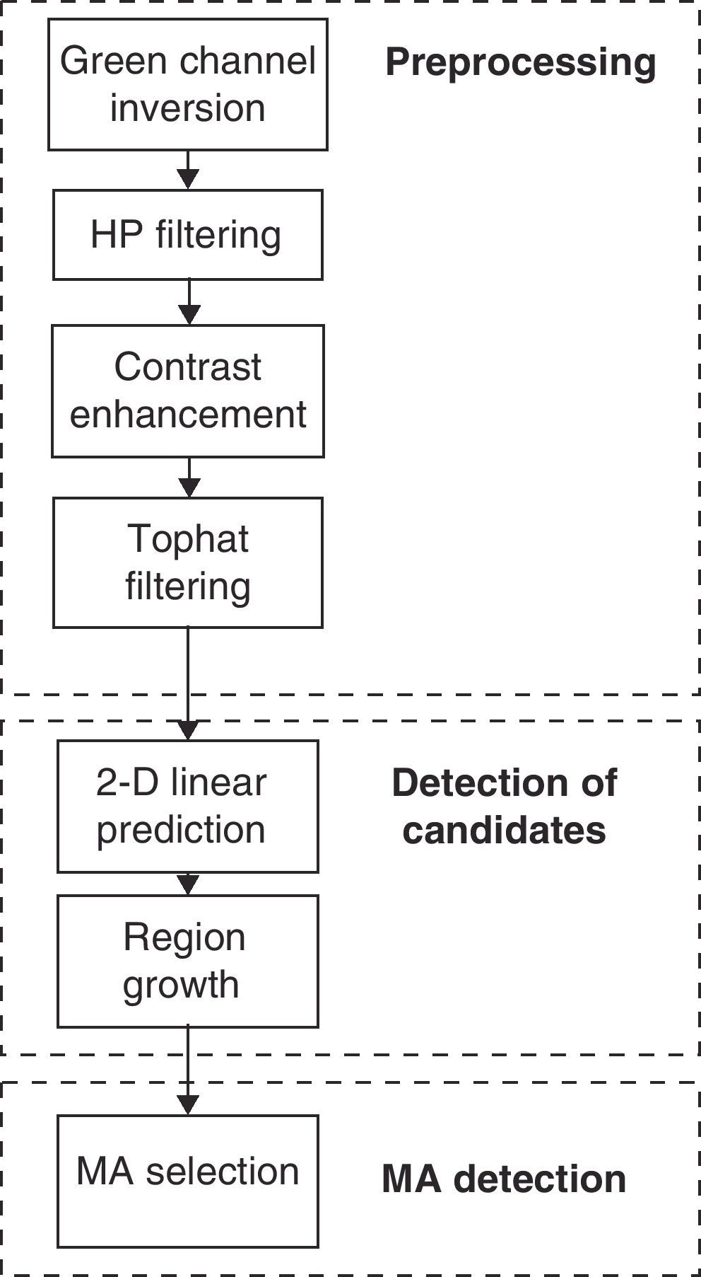
MethodThe designed and developed tool consists of three stages of processing: (1) Obtaining of the basic image of eye with the retinal camera, inverted image on the green channel, and a high-pass filter of the image. This phase enhances the microaneurysms. (2) Detection of the candidates for microaneurysms, by means of an adaptive prediction filter and regions growth. (3) Selection, among the candidates, of whom microaneurysms must be considered to fulfil the criteria of circular shape, high intensity in the inverted green channel and contrasts with respect to the surrounding pixels.
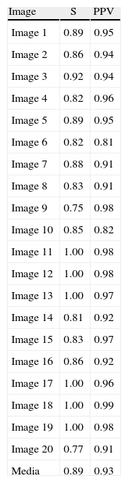
ResultsWe selected to 20 retinal photographs of good quality and dimensions 600×600 pixels from patients with nonproliferative diabetic retinopathy. The ophthalmologists detected 297 microaneurysms in these images. The tool for automatic detection correctly located 252 microaneurysms, with a mean sensitivity of 89% and a false positives rate of 93%.
ConclusionsThe results obtained seem to indicate that the tool developed will be very useful for its potential use in screening programs in primary care centres. On the other hand, more work is needed on the algorithm to decrease the rate of false positives.
Presentamos el desarrollo de una herramienta para la detección automática de microaneurismas y su evaluación clínica. El propósito de esta herramienta es facilitar el diagnóstico de lesiones diabéticas en programas generales de detección.
MétodoLa herramienta diseñada y desarrollada consta de tres etapas de procesamiento: 1) Obtención de la imagen de fondo de ojo con el retinógrafo, inversión del canal verde y filtrado paso de alta de la imagen. Esta fase realza los microaneurismas. 2) Detección de los candidatos a microaneurismas, mediante un filtrado de predicción adaptativo y un crecimiento de regiones. 3) Selección, de entre los candidatos, de los que deben considerarse microaneurismas por cumplir con los criterios de: forma circular, intensidad alta en el canal verde invertido y contraste respecto a los píxeles de alrededor.
ResultadosSe seleccionaron 20 retinografías de buena calidad y dimensiones 600×600 píxeles de pacientes con retinopatía diabética no proliferante. Los oftalmólogos detectaron un total de 297 microaneurismas en estas imágenes. La herramienta de detección automática localizó adecuadamente 252 microaneurismas, con una sensibilidad media del 89% y una tasa de falsos positivos del 93%.
ConclusionesLos resultados obtenidos parecen indicar que la herramienta desarrollada podría ser muy útil para su potencial utilización en programas de detección en los centros de asistencia primaria. Por otro lado, es necesario seguir trabajando en el algoritmo para disminuir la tasa de falsos positivos.