Congenital malformations and acquired lesions of the inner ear are characterised by small structural changes in this region. In recent decades, treatment options have improved considerably. At the same time, there has been a great advancement in diagnostic methods, obtaining high-resolution labyrinth images.
Currently, we use a 64-multislice computed tomography scanner in spiral mode (Brilliance 64 Phillips, Eindhoven, the Netherlands), with an overlap of 0.66mm and an interval of 0.33mm, 120kV and 300mA. The magnetic resonance images were taken with Signa HDxt 1.5 and 3.0T units (GE Healthcare, Waukesha, WI, USA).
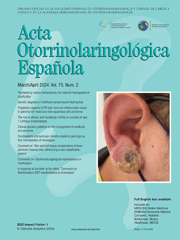
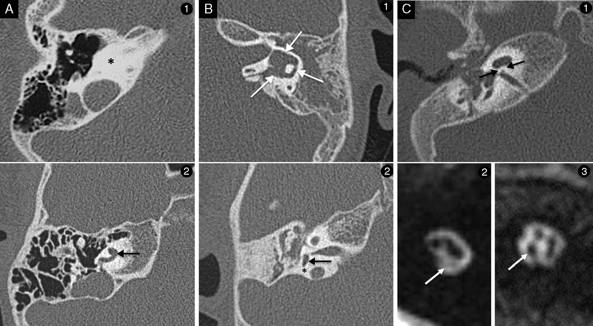
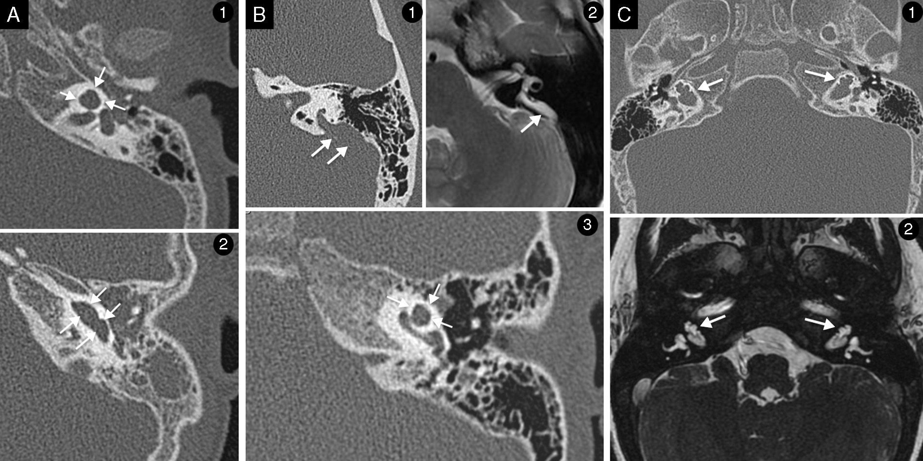
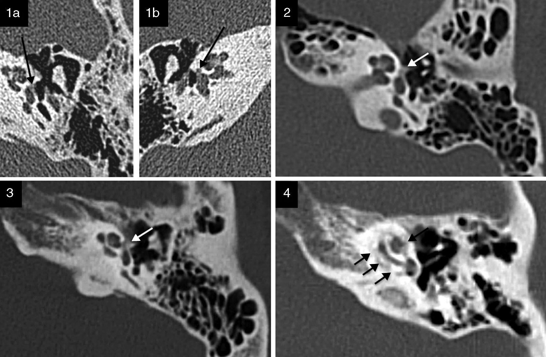
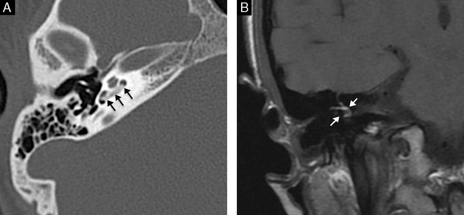
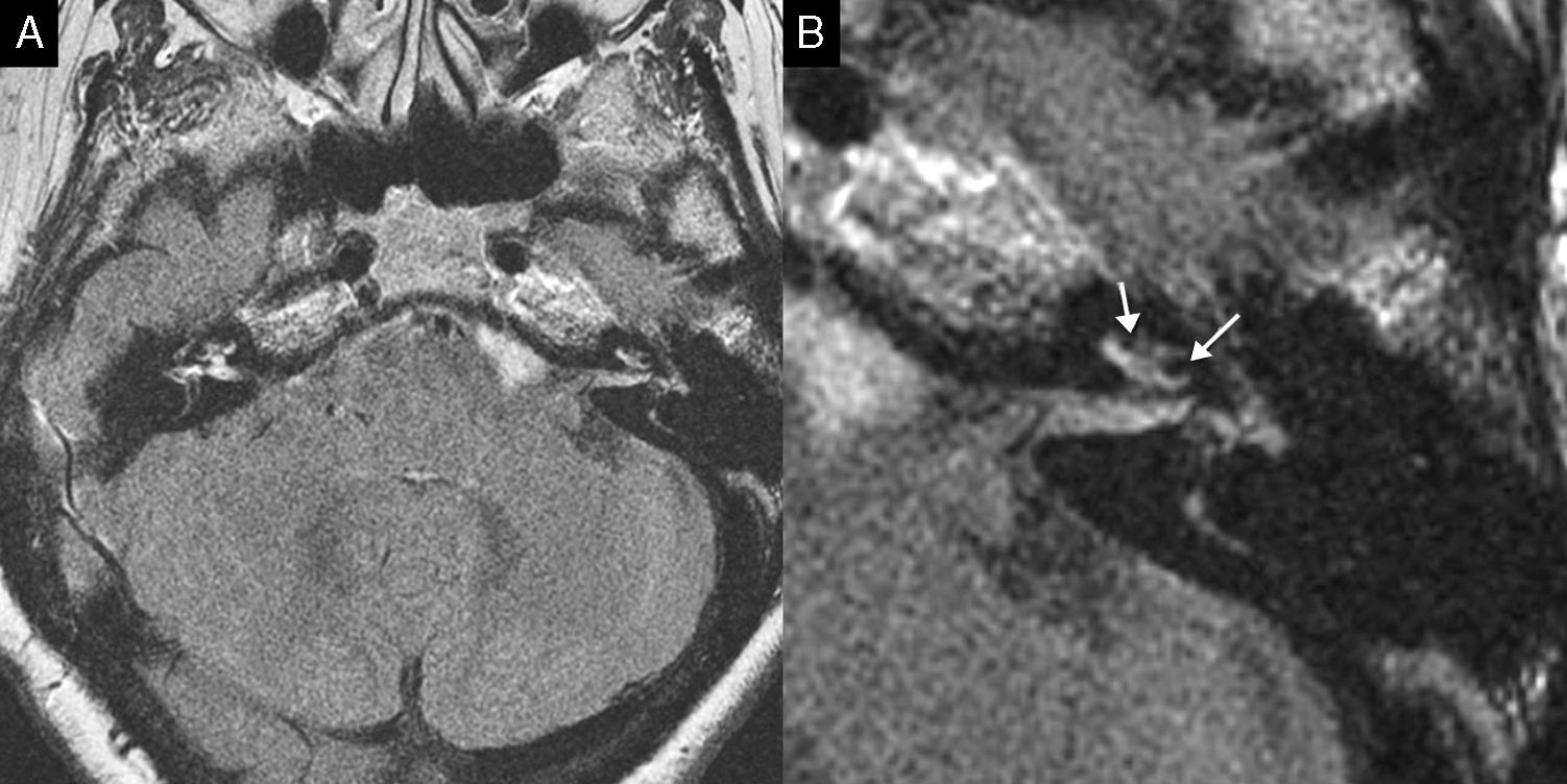
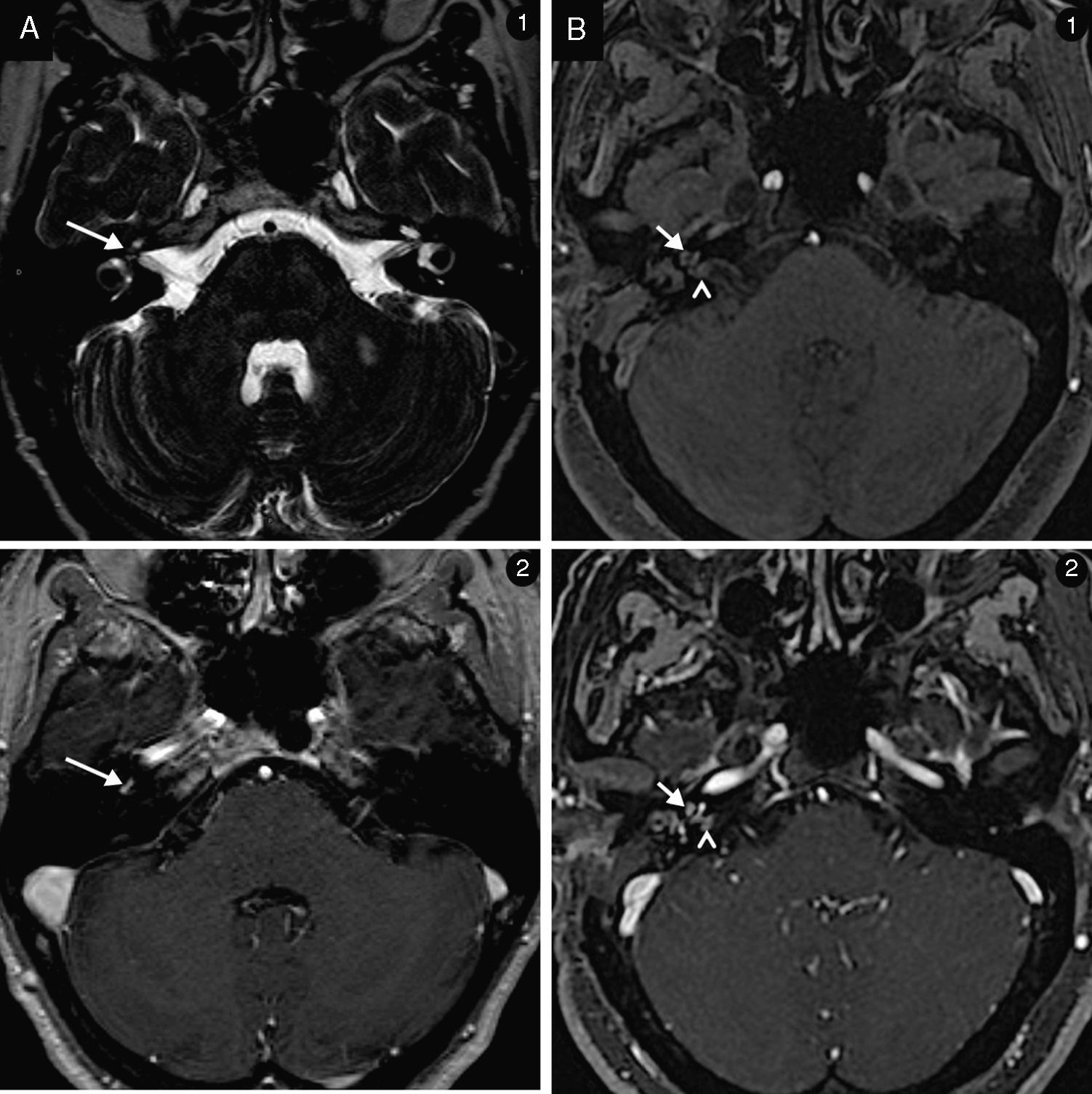
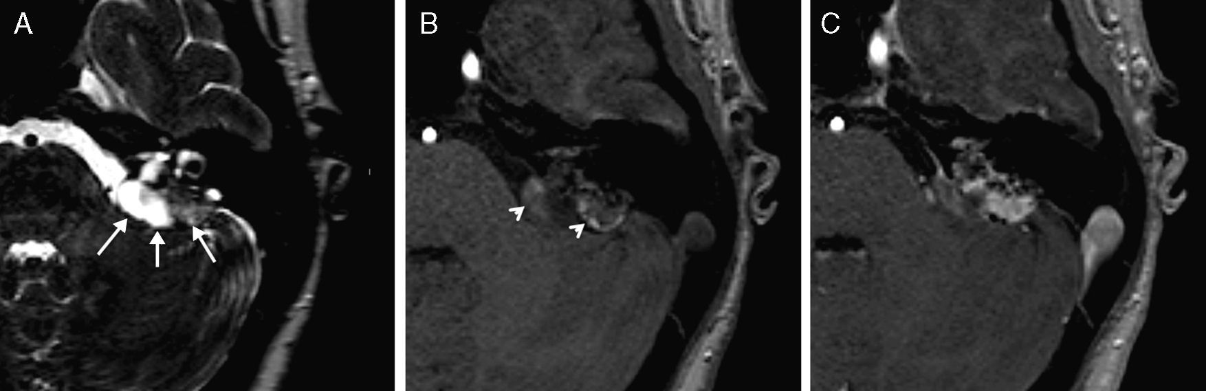
We reviewed the radiological features of the lesions affecting the inner ear. They are classified as congenital (labyrinth malformation and statoacoustic nerve deficiencies) or acquired (otospongiosis, labyrinthitis, Ménière's disease, inner ear haemorrhage, intralabyrinthine schwannoma and endolymphatic sac tumour).
ConclusionMagnetic resonance imaging and computed tomography play an essential role in diagnosing patients with inner ear pathology. The technique selected should be chosen depending on the clinical setting. In a generic way, tomography is the method of choice for the study of traumatic pathology or otospongiosis. When tumour or inflammatory pathology is suspected, magnetic resonance is superior. In cases of congenital malformation, both techniques are complementary.
Las malformaciones congénitas y las lesiones adquiridas del oído interno se caracterizan por pequeños cambios estructurales de esta región. En las últimas décadas, las opciones terapéuticas han mejorado considerablemente, y paralelamente se ha producido un gran avance en los métodos diagnósticos, consiguiendo imágenes de alta resolución del laberinto.
Actualmente se utiliza una tomografía computerizada multicorte de 64 detectores (Brilliance 64 Phillips, Eindhoven, the Netherlands), un espesor de adquisición de 0,66 y un intervalo de 0,33mm, 120KV y 300mA. Las imágenes de resonancia magnética proceden de los equipos Signa HDxt 1.5 y 3.0T (GE Healthcare, Waukesha, WI, USA).
Se realiza una revisión de las características radiológicas de las lesiones que afectan al oído interno que son clasificadas según su origen en congénitas (malformaciones del laberinto y deficiencias de los nervios estatoacústicos) o adquiridas (otoespongiosis, laberintitis, hemorragia del oído interno, enfermedad de Menière, schwannoma intralaberíntico, tumour del saco endolinfático).
ConclusionesLa resonancia magnética y la tomografía computerizada juegan un papel fundamental en el diagnóstico de pacientes con patología del oído interno. La técnica de elección debe ser escogida en función del escenario clínico. De forma genérica, para el estudio de patología traumática u otoespongiosis la tomografia es el método de elección. Ante sospecha de patología tumoral o inflamatoria la resonancia se muestra superior. Para el estudio de patología malformativa ambas técnicas son complementarias.